1JRG image
Deposition Date 2001-08-13
Release Date 2002-06-19
Last Version Date 2024-11-06
Entry Detail
PDB ID:
1JRG
Keywords:
Title:
Crystal Structure of the R3 form of Pectate Lyase A, Erwinia chrysanthemi
Biological Source:
Source Organism:
Host Organism:
Method Details:
Experimental Method:
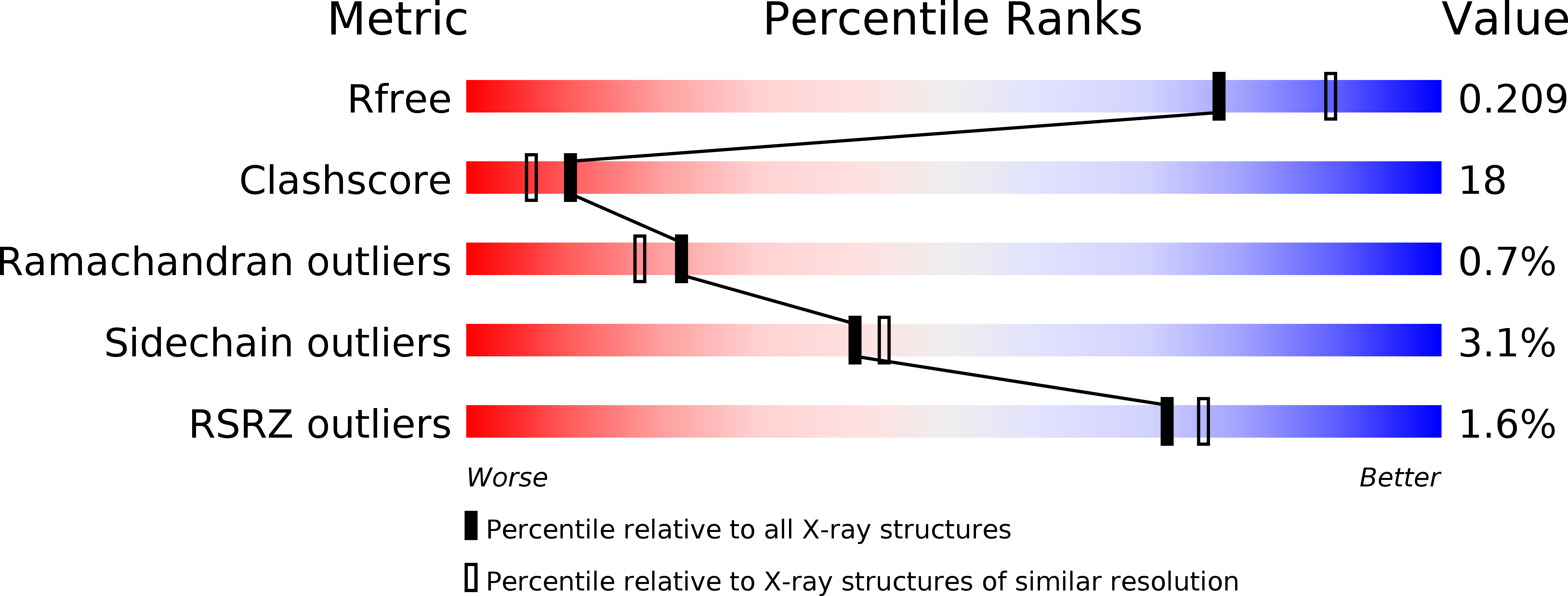
Resolution:
2.10 Å
R-Value Free:
0.21
R-Value Work:
0.16
R-Value Observed:
0.16
Space Group:
H 3
Macromolecular Entities
Polymer Type:polypeptide(L)
Molecule:Pectate lyase
Chain IDs:A, B
Chain Length:361
Number of Molecules:2
Biological Source:Erwinia chrysanthemi
Ligand Molecules
Primary Citation
Structure of pectate lyase A: comparison to other isoforms.
Acta Crystallogr.,Sect.D 58 1008 1015 (2002)
PMID: 12037303 DOI: 10.1107/S0907444902005851

Abstact

Pectate lyase A is a virulence factor secreted by the plant-pathogenic bacteria Erwinia chrysanthemi. The enzyme cleaves the glycosidic bond of pectate polymers by a calcium-dependent beta-elimination mechanism. The crystal structure of pectate lyase A from E. chrysanthemi EC16 has been determined in two crystal forms, monoclinic C2 to 1.8 A and rhombohedral R3 to 2.1 A. The protein structure is compared with two other pectate lyase isoforms from E. chrysanthemi EC16, pectate lyase C and pectate lyase E. Pectate lyase A is unique as it is the only acidic pectate lyase and has end products that are significantly more varied in length in comparison to those of the other four major pectate lyase isozymes. Differences and similarities in polypeptide trace, size and volume of the active-site groove and surface electrostatics are discussed.

Legend

Protein

Chemical

Disease

Primary Citation of related structures