Deposition Date
2001-08-04
Release Date
2002-11-06
Last Version Date
2024-10-09
Entry Detail
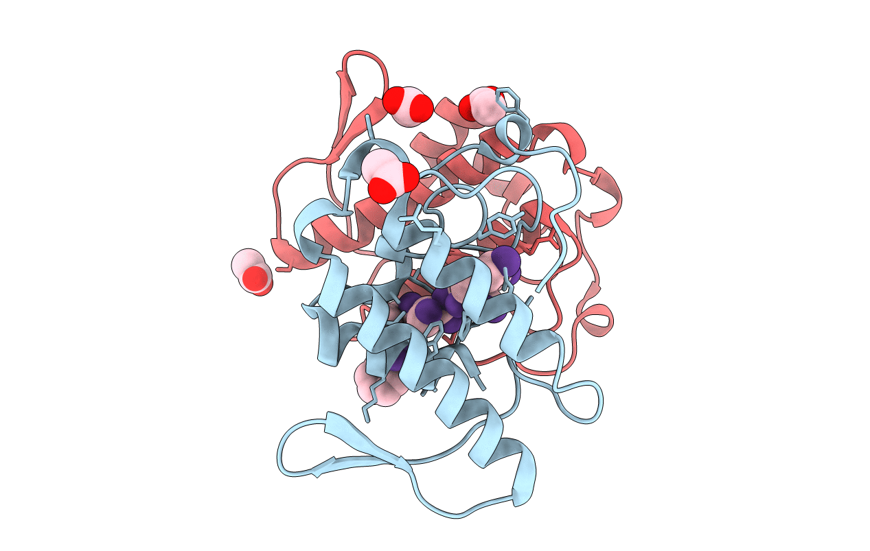
PDB ID:
1JQ9
Keywords:
Title:
Crystal structure of a complex formed between phospholipase A2 from Daboia russelli pulchella and a designed pentapeptide Phe-Leu-Ser-Tyr-Lys at 1.8 resolution
Biological Source:
Source Organism(s):
Daboia russellii pulchella (Taxon ID: 97228)
Method Details:
Experimental Method:
Resolution:
1.80 Å
R-Value Free:
0.22
R-Value Work:
0.20
R-Value Observed:
0.20
Space Group:
C 2 2 21