1JPE image
Deposition Date 2001-08-02
Release Date 2002-09-25
Last Version Date 2024-10-30
Entry Detail
PDB ID:
1JPE
Title:
Crystal structure of DsbD-alpha; the N-terminal domain of DsbD
Biological Source:
Source Organism:
Escherichia coli (Taxon ID: 562)
Host Organism:
Method Details:
Experimental Method:
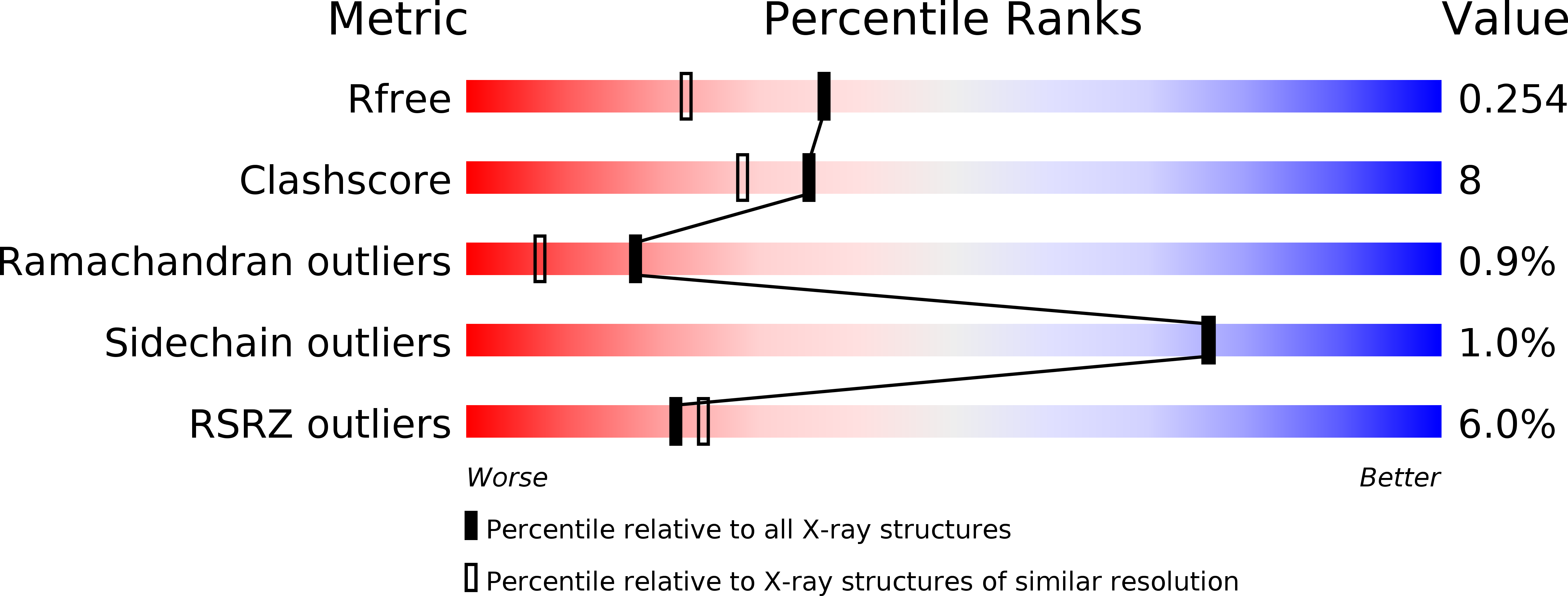
Resolution:
1.90 Å
R-Value Free:
0.25
R-Value Work:
0.21
R-Value Observed:
0.22
Space Group:
C 2 2 21
Macromolecular Entities
Structures with similar UniProt ID
Protein Blast
Polymer Type:polypeptide(L)
Molecule:DsbD-alpha
Gene (Uniprot):dsbD
Chain IDs:A
Chain Length:151
Number of Molecules:1
Biological Source:Escherichia coli
Primary Citation
The disulfide bond isomerase DsbC is activated by an immunoglobulin-fold thiol oxidoreductase: crystal structure of the DsbC-DsbD alpha complex.
Embo J. 21 4774 4784 (2002)
PMID: 12234918 DOI: 10.1093/emboj/cdf489

Abstact

The Escherichia coli disulfide bond isomerase DsbC rearranges incorrect disulfide bonds during oxidative protein folding. It is specifically activated by the periplasmic N-terminal domain (DsbDalpha) of the transmembrane electron transporter DsbD. An intermediate of the electron transport reaction was trapped, yielding a covalent DsbC-DsbDalpha complex. The 2.3 A crystal structure of the complex shows for the first time the specific interactions between two thiol oxidoreductases. DsbDalpha is a novel thiol oxidoreductase with the active site cysteines embedded in an immunoglobulin fold. It binds into the central cleft of the V-shaped DsbC dimer, which assumes a closed conformation on complex formation. Comparison of the complex with oxidized DsbDalpha reveals major conformational changes in a cap structure that regulates the accessibility of the DsbDalpha active site. Our results explain how DsbC is selectively activated by DsbD using electrons derived from the cytoplasm.

Legend

Protein

Chemical

Disease

Primary Citation of related structures
Feedback Form
Name
Email
Institute
Feedback