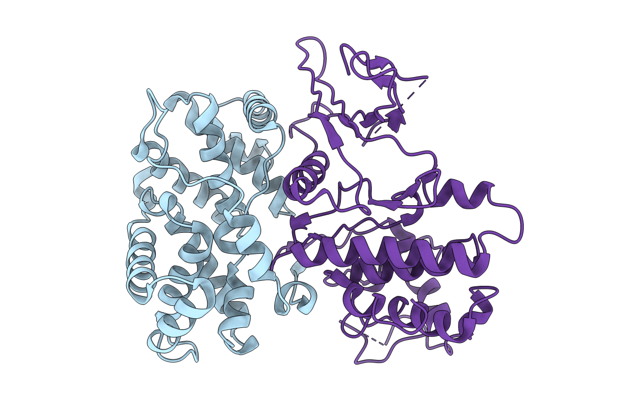
1JOW image
Entry Detail
PDB ID:
1JOW
Title:
Crystal structure of a complex of human CDK6 and a viral cyclin
Biological Source:
PDB Version:
Deposition Date:
2001-07-31
Release Date:
2002-02-27
Method Details:
Experimental Method:
Resolution:
3.10 Å
R-Value Free:
0.32
R-Value Work:
0.26
Space Group:
P 65 2 2
Macromolecular Entities
Polymer Type:polypeptide(L)
Description:CYCLIN HOMOLOG
Chain IDs:A
Chain Length:254
Number of Molecules:1
Biological Source:Saimiriine herpesvirus 2
Polymer Type:polypeptide(L)
Description:CELL DIVISION PROTEIN KINASE 6
Chain IDs:B
Chain Length:308
Number of Molecules:1
Biological Source:Homo sapiens
Ligand Molecules
Primary Citation
Structural basis for CDK6 activation by a virus-encoded cyclin.
Nat.Struct.Biol. 9 177 181 (2002)
PMID: 11828325

Abstact

Cyclin from herpesvirus saimiri (Vcyclin) preferentially forms complexes with cyclin-dependent kinase 6 (CDK6) from primate host cells. These complexes show higher kinase activity than host cell CDKs in complex with cellular cyclins and are resistant to cyclin-dependent inhibitory proteins (CDKIs). The crystal structure of human CDK6--Vcyclin in an active state was determined to 3.1 A resolution to better understand the structural basis of CDK6 activation by viral cyclins. The unphosphorylated CDK6 in complex with Vcyclin has many features characteristic of cyclinA-activated, phosphorylated CDK2. There are, however, differences in the conformation at the tip of the T-loop and its interactions with Vcyclin. Residues in the N-terminal extension of Vcyclin wrap around the tip of the CDK6 T-loop and form a short beta-sheet with the T-loop backbone. These interactions lead to a 20% larger buried surface in the CDK6--Vcyclin interface than in the CDK2--cyclinA complex and are probably largely responsible for the specificity of Vcyclin for CDK6 and resistance of the complex to inhibition by INK-type CDKIs.

Legend

Protein

Chemical

Disease

Primary Citation of related structures