
Deposition Date
2001-07-31
Release Date
2001-08-10
Last Version Date
2024-10-30
Entry Detail
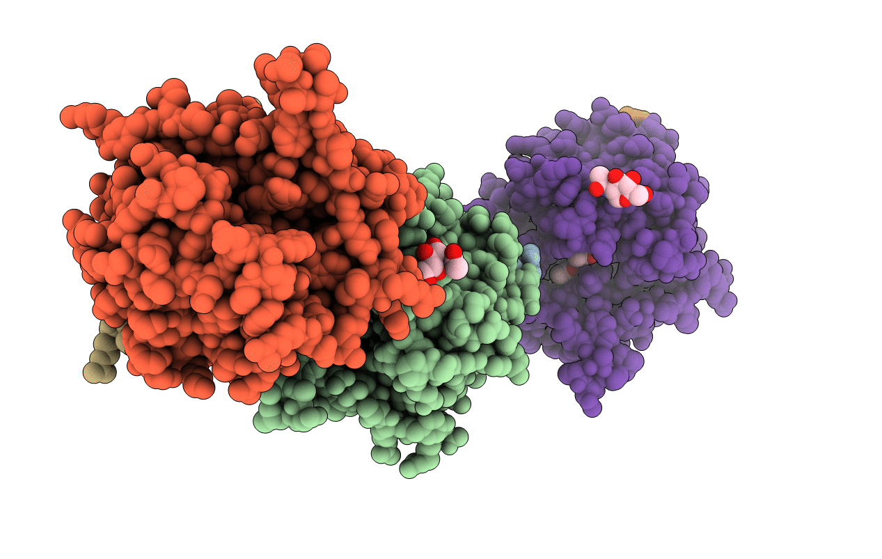
PDB ID:
1JOU
Keywords:
Title:
Crystal Structure of Native S195A Thrombin with an Unoccupied Active Site
Biological Source:
Source Organism(s):
Homo sapiens (Taxon ID: 9606)
Expression System(s):
Method Details:
Experimental Method:
Resolution:
1.80 Å
R-Value Free:
0.24
R-Value Work:
0.22
Space Group:
P 1 21 1
