1JMO image
Deposition Date 2001-07-19
Release Date 2002-08-30
Last Version Date 2024-10-30
Entry Detail
PDB ID:
1JMO
Keywords:
Title:
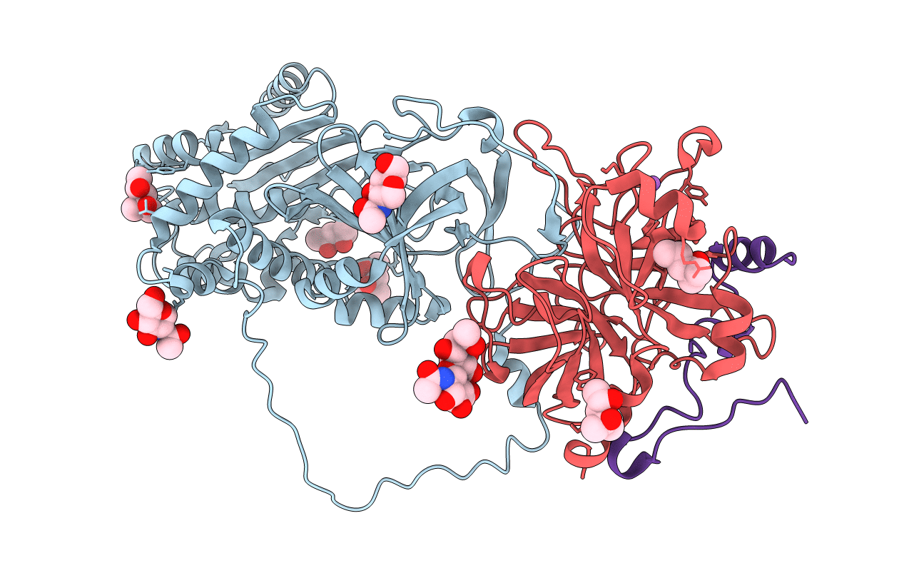
Crystal Structure of the Heparin Cofactor II-S195A Thrombin Complex
Biological Source:
Source Organism:
Homo sapiens (Taxon ID: 9606)
Host Organism:
Method Details:
Experimental Method:
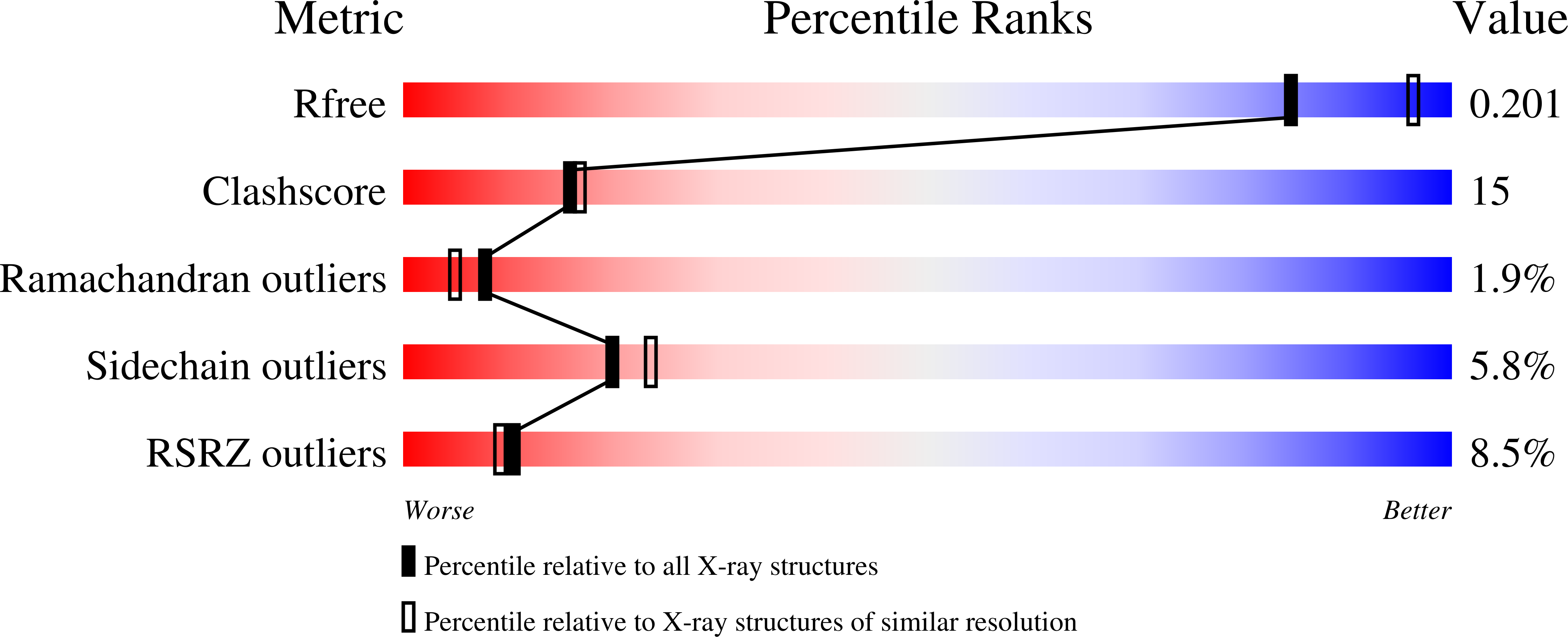
Resolution:
2.20 Å
R-Value Free:
0.21
R-Value Work:
0.20
Space Group:
P 61
Macromolecular Entities
Polymer Type:polypeptide(L)
Molecule:HEPARIN COFACTOR II
Gene (Uniprot):SERPIND1
Chain IDs:C (auth: A)
Chain Length:480
Number of Molecules:1
Biological Source:Homo sapiens
Polymer Type:polypeptide(L)
Molecule:Thrombin, heavy chain
Gene (Uniprot):F2
Mutations:S195A
Chain IDs:B (auth: H)
Chain Length:260
Number of Molecules:1
Biological Source:Homo sapiens
Polymer Type:polypeptide(L)
Molecule:Thrombin, light chain
Gene (Uniprot):F2
Chain IDs:A (auth: L)
Chain Length:48
Number of Molecules:1
Biological Source:Homo sapiens
Modified Residue
Compound ID Chain ID Parent Comp ID Details 2D Image
ASN B ASN GLYCOSYLATION SITE
TYS C TYR O-SULFO-L-TYROSINE
Primary Citation
Crystal structures of native and thrombin-complexed heparin cofactor II reveal a multistep allosteric mechanism.
Proc.Natl.Acad.Sci.USA 99 11079 11084 (2002)
PMID: 12169660 DOI: 10.1073/pnas.162232399

Abstact

The serine proteases sequentially activated to form a fibrin clot are inhibited primarily by members of the serpin family, which use a unique beta-sheet expansion mechanism to trap and destroy their targets. Since the discovery that serpins were a family of serine protease inhibitors there has been controversy as to the role of conformational change in their mechanism. It now is clear that protease inhibition depends entirely on rapid serpin beta-sheet expansion after proteolytic attack. The regulatory advantage afforded by the conformational mobility of serpins is demonstrated here by the structures of native and S195A thrombin-complexed heparin cofactor II (HCII). HCII inhibits thrombin, the final protease of the coagulation cascade, in a glycosaminoglycan-dependent manner that involves the release of a sequestered hirudin-like N-terminal tail for interaction with thrombin. The native structure of HCII resembles that of native antithrombin and suggests an alternative mechanism of allosteric activation, whereas the structure of the S195A thrombin-HCII complex defines the molecular basis of allostery. Together, these structures reveal a multistep allosteric mechanism that relies on sequential contraction and expansion of the central beta-sheet of HCII.

Legend

Protein

Chemical

Disease

Primary Citation of related structures