1JKZ image
Deposition Date 2001-07-13
Release Date 2002-02-06
Last Version Date 2024-10-30
Entry Detail
PDB ID:
1JKZ
Title:
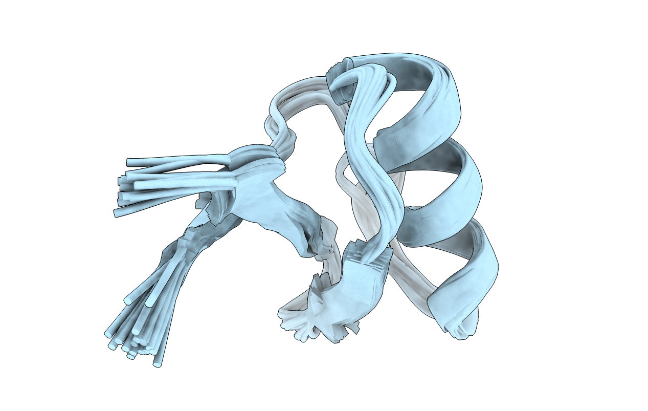
NMR Solution Structure of Pisum sativum defensin 1 (Psd1)
Biological Source:
Source Organism:
Pisum sativum (Taxon ID: 3888)
Method Details:
Experimental Method:
Conformers Calculated:
100
Conformers Submitted:
20
Selection Criteria:
structures with the lowest energy
Macromolecular Entities
Polymer Type:polypeptide(L)
Molecule:DEFENSE-RELATED PEPTIDE 1
Chain IDs:A
Chain Length:46
Number of Molecules:1
Biological Source:Pisum sativum
Ligand Molecules
Primary Citation
Solution structure of Pisum sativum defensin 1 by high resolution NMR: plant defensins, identical backbone with different mechanisms of action.
J.Mol.Biol. 315 749 757 (2002)
PMID: 11812144 DOI: 10.1006/jmbi.2001.5252

Abstact

Pisum sativum defensin 1 (Psd1) is a 46 amino acid residue plant defensin isolated from seeds of pea. The three-dimensional structure in solution of Psd1 was determined by two-dimensional NMR data recorded at 600 MHz. Experimental restraints were used for structure calculation using CNS and torsion-angle molecular dynamics. The 20 lowest energy structures were selected and further subjected to minimization, giving a root-mean-square deviation of 0.78(+/- 0.22) A in the backbone and 1.91(+/-0.60) A for over all atoms of the molecule. The protein has a globular fold with a triple-stranded antiparalell beta-sheet and an alpha-helix (from residue Asn17 to Leu27). Psd1 presents the so called "cysteine stabilized alpha/beta motif" and presents identical three-dimensional topology in the backbone with other defensins and neurotoxins. Comparison of the electrostatic surface potential among proteins with high three-dimensional (selected using the softwares TOP and DALI) topology gave insights into the mode of action of Psd1. The surface topologies between proteins that present antifungal activity or sodium channel inhibiting activity are different. On the other hand the surface topology presents several common features with potassium channel inhibitors, suggesting that Psd1 presents this activity. Other common features with potassium channel inhibitors were found including the presence of a lysine residue essential for inhibitory activity. The identity of Psd1 in primary sequence is not enough to infer a mechanism of action, in contrast with the strategy proposed here.

Legend

Protein

Chemical

Disease

Primary Citation of related structures