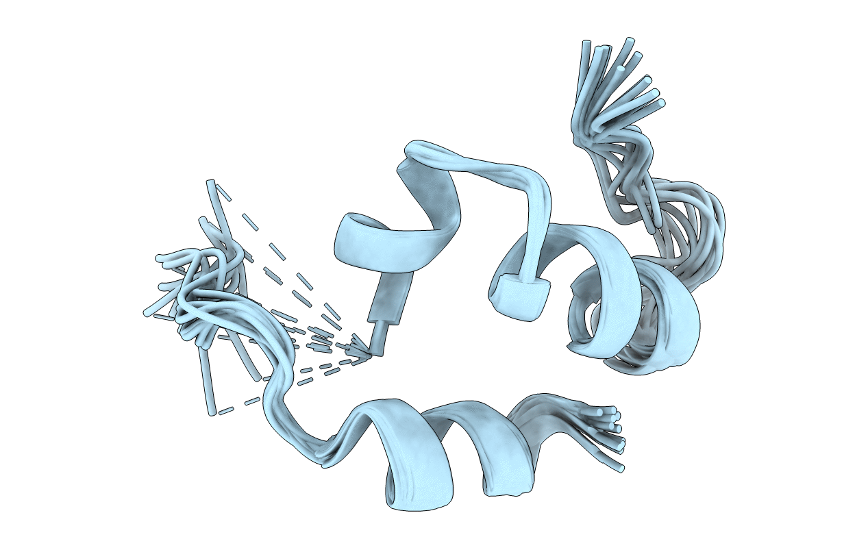
1JJS image
Deposition Date 2001-07-09
Release Date 2001-10-03
Last Version Date 2024-05-22
Entry Detail
PDB ID:
1JJS
Keywords:
Title:
NMR Structure of IBiD, A Domain of CBP/p300
Biological Source:
Source Organism:
Mus musculus (Taxon ID: 10090)
Host Organism:
Method Details:
Experimental Method:
Conformers Calculated:
50
Conformers Submitted:
12
Selection Criteria:
structures with the least restraint violations
Macromolecular Entities
Structures with similar UniProt ID
Protein Blast
Polymer Type:polypeptide(L)
Molecule:CREB-BINDING PROTEIN
Gene (Uniprot):Crebbp
Chain IDs:A
Chain Length:50
Number of Molecules:1
Biological Source:Mus musculus
Ligand Molecules
Primary Citation
A small domain of CBP/p300 binds diverse proteins: solution structure and functional studies.
Mol.Cell 8 581 590 (2001)
PMID: 11583620 DOI: 10.1016/S1097-2765(01)00333-1

Abstact

The transcriptional coactivators CBP and p300 are critical regulators of metazoan gene expression. They associate with many different DNA-bound transcription factors through small, conserved domains. We have identified a compactly folded 46 residue domain in CBP and p300, the IRF-3 binding domain (IBiD), and we have determined its structure by NMR. It has a helical framework containing an apparently flexible polyglutamine loop that participates in ligand binding. Spectroscopic data indicate that induced folding accompanies association of IBiD with its partners, which exhibit no evident sequence similarities. We demonstrate the significance both in vitro and in vivo of interactions between IBiD and a number of diverse partners. Thus, IBiD is an important contributor to signal integration by CBP and p300.

Legend

Protein

Chemical

Disease

Primary Citation of related structures