
Deposition Date
2001-07-03
Release Date
2002-07-03
Last Version Date
2024-02-07
Entry Detail
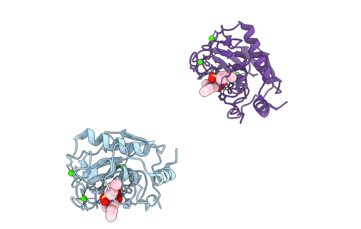
PDB ID:
1JIZ
Keywords:
Title:
Crystal Structure Analysis of human Macrophage Elastase MMP-12
Biological Source:
Source Organism(s):
Homo sapiens (Taxon ID: 9606)
Expression System(s):
Method Details:
Experimental Method:
Resolution:
2.60 Å
R-Value Free:
0.25
R-Value Work:
0.19
Space Group:
I 2 2 2
