Deposition Date
2001-06-28
Release Date
2002-02-08
Last Version Date
2023-08-16
Entry Detail
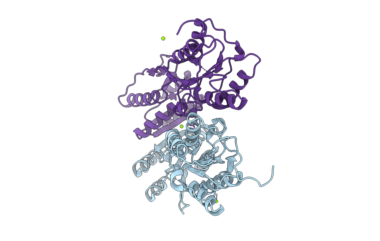
PDB ID:
1JHZ
Keywords:
Title:
Purine Repressor Mutant Corepressor Binding Domain Structure
Biological Source:
Source Organism:
Escherichia coli (Taxon ID: 562)
Host Organism:
Method Details:
Experimental Method:
Resolution:
2.40 Å
R-Value Observed:
0.17
Space Group:
P 1 21 1