1JHN image
Deposition Date 2001-06-28
Release Date 2001-10-10
Last Version Date 2024-11-13
Entry Detail
PDB ID:
1JHN
Keywords:
Title:
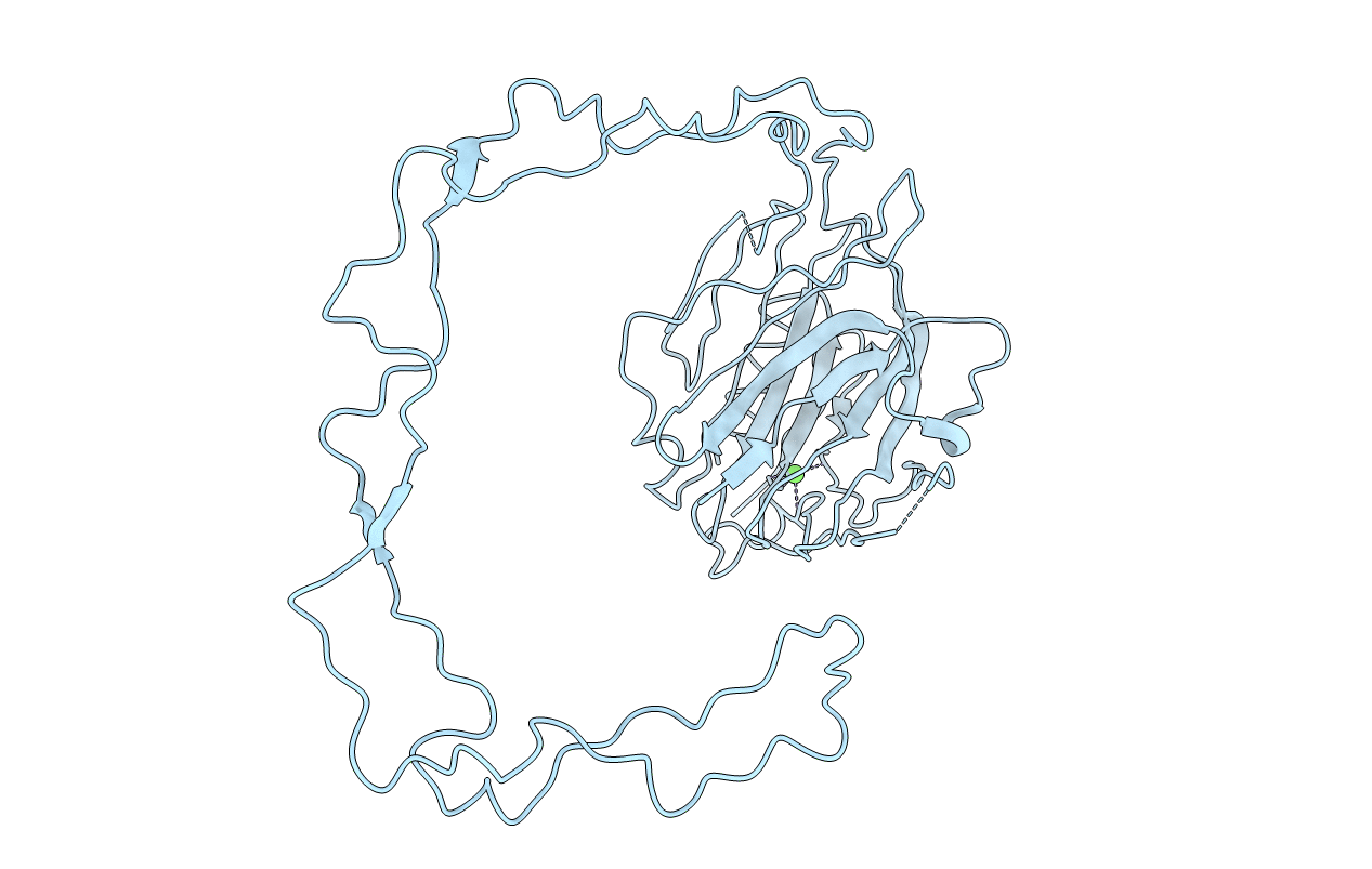
Crystal Structure of the Lumenal Domain of Calnexin
Biological Source:
Source Organism:
Host Organism:
Method Details:
Experimental Method:
Resolution:
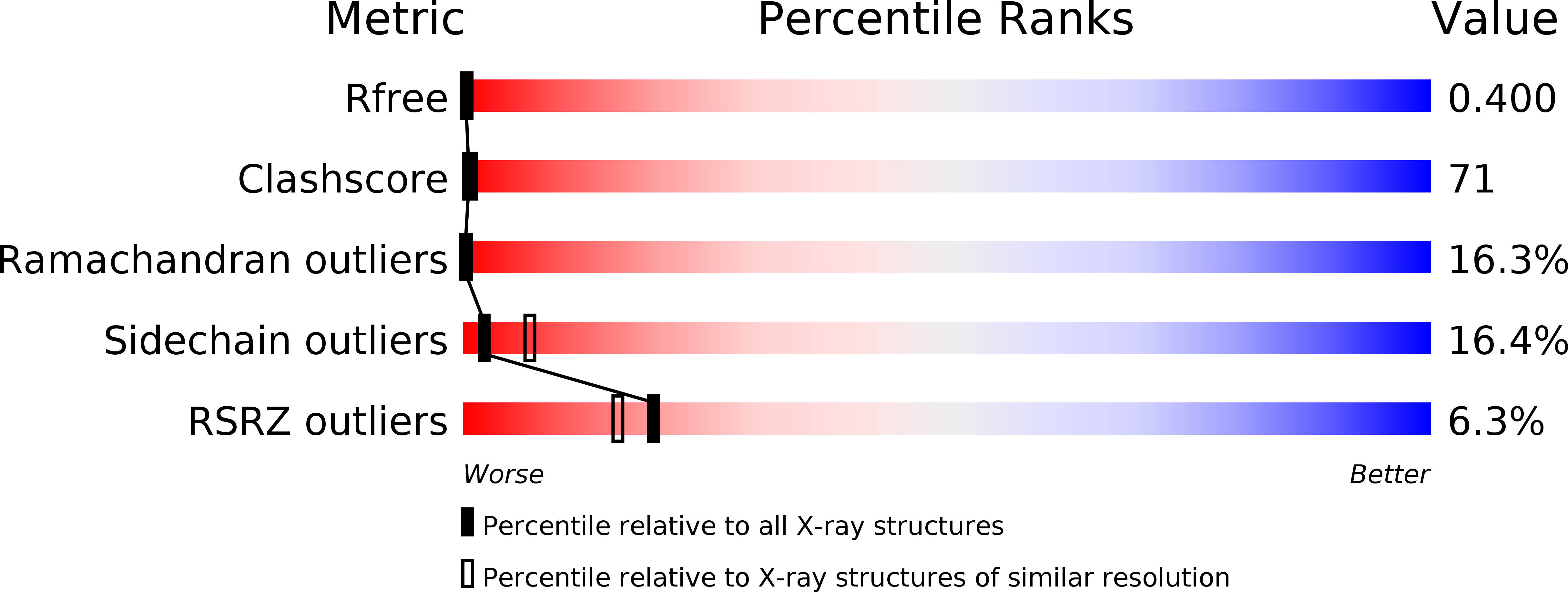
2.90 Å
R-Value Free:
0.37
R-Value Work:
0.32
Space Group:
P 43 21 2
Macromolecular Entities
Polymer Type:polypeptide(L)
Molecule:calnexin
Gene (Uniprot):CANX
Chain IDs:A
Chain Length:424
Number of Molecules:1
Biological Source:Canis lupus familiaris
Ligand Molecules
Primary Citation
The Structure of calnexin, an ER chaperone involved in quality control of protein folding.
Mol.Cell 8 633 644 (2001)
PMID: 11583625 DOI: 10.1016/S1097-2765(01)00318-5

Abstact

The three-dimensional structure of the lumenal domain of the lectin-like chaperone calnexin determined to 2.9 A resolution reveals an extended 140 A arm inserted into a beta sandwich structure characteristic of legume lectins. The arm is composed of tandem repeats of two proline-rich sequence motifs which interact with one another in a head-to-tail fashion. Identification of the ligand binding site establishes calnexin as a monovalent lectin, providing insight into the mechanism by which the calnexin family of chaperones interacts with monoglucosylated glycoproteins.

Legend

Protein

Chemical

Disease

Primary Citation of related structures