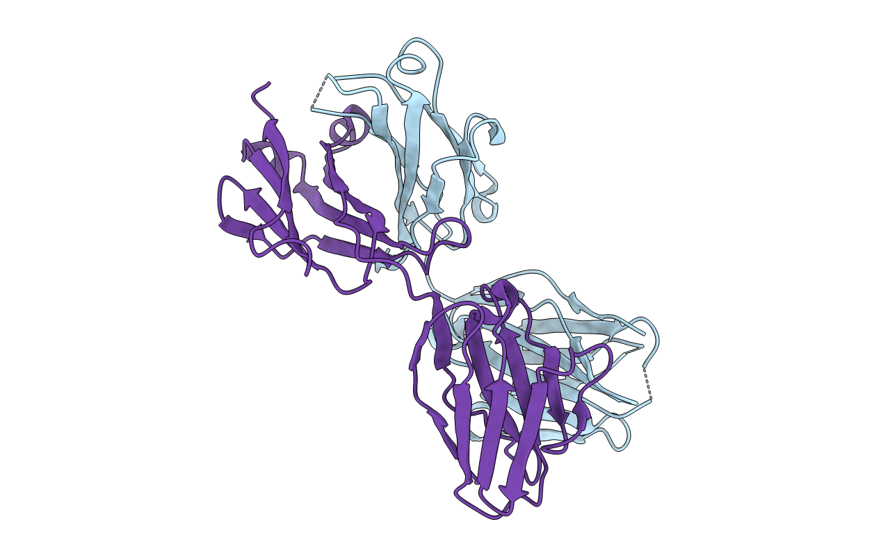
1JHK image
Deposition Date 2001-06-28
Release Date 2001-10-10
Last Version Date 2024-11-06
Entry Detail
PDB ID:
1JHK
Keywords:
Title:
Crystal structure of the anti-estradiol antibody 57-2
Biological Source:
Source Organism:
Mus musculus (Taxon ID: 10090)
Host Organism:
Method Details:
Experimental Method:
Resolution:
2.51 Å
R-Value Free:
0.26
R-Value Work:
0.19
R-Value Observed:
0.23
Space Group:
P 21 21 21
Macromolecular Entities
Structures with similar UniProt ID
Protein Blast
Polymer Type:polypeptide(L)
Molecule:Ig gamma-1-chain
Chain IDs:B (auth: H)
Chain Length:218
Number of Molecules:1
Biological Source:Mus musculus
Structures with similar UniProt ID
Protein Blast
Polymer Type:polypeptide(L)
Molecule:Ig kappa-chain
Chain IDs:A (auth: L)
Chain Length:214
Number of Molecules:1
Biological Source:Mus musculus
Primary Citation
Crystal structure of a recombinant anti-estradiol Fab fragment in complex with 17beta -estradiol.
J.Biol.Chem. 276 36687 36694 (2001)
PMID: 11451948 DOI: 10.1074/jbc.M102367200

Abstact

The crystal structure of a Fab fragment of an anti-17beta-estradiol antibody 57-2 was determined in the absence and presence of the steroid ligand, 17beta-estradiol (E2), at 2.5 and 2.15-A resolutions, respectively. The antibody binds the steroid in a deep hydrophobic pocket formed at the interface between the variable domains. No major structural rearrangements take place upon ligand binding; however, a large part of the heavy chain variable domain near the binding pocket is unusually flexible and is partly stabilized when the steroid is bound. The nonpolar steroid skeleton of E2 is recognized by a number of hydrophobic interactions, whereas the two hydroxyl groups of E2 are hydrogen-bonded to the protein. Especially, the 17-hydroxyl group of E2 is recognized by an intricate hydrogen bonding network in which the 17-hydroxyl itself forms a rare four-center hydrogen bond with three polar amino acids; this hydrogen bonding arrangement accounts for the low cross-reactivity of the antibody with other estrogens such as estrone. The CDRH3 loop plays a prominent role in ligand binding. All the complementarity-determining regions of the light chain make direct contacts with the steroid, even CDRL2, which is rarely directly involved in the binding of haptens.

Legend

Protein

Chemical

Disease

Primary Citation of related structures