1JHB image
Deposition Date 1998-02-17
Release Date 1998-08-26
Last Version Date 2024-05-22
Entry Detail
PDB ID:
1JHB
Title:
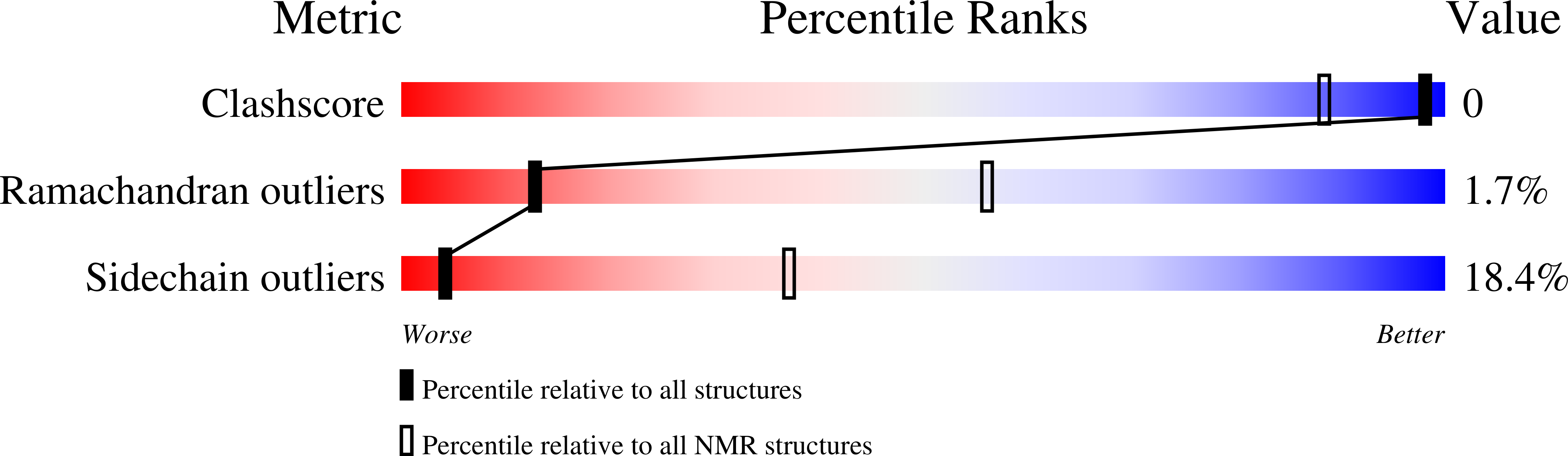
HUMAN GLUTAREDOXIN IN FULLY REDUCED FORM, NMR, 20 STRUCTURES
Biological Source:
Source Organism:
Homo sapiens (Taxon ID: 9606)
Host Organism:
Method Details:
Experimental Method:
Conformers Calculated:
50
Conformers Submitted:
20
Selection Criteria:
LEAST RESTRAINT VIOLATION
Macromolecular Entities
Structures with similar UniProt ID
Protein Blast
Polymer Type:polypeptide(L)
Molecule:GLUTAREDOXIN
Gene (Uniprot):GLRX
Chain IDs:A
Chain Length:106
Number of Molecules:1
Biological Source:Homo sapiens
Ligand Molecules
Primary Citation
The NMR solution structure of human glutaredoxin in the fully reduced form.
J.Mol.Biol. 280 687 701 (1998)
PMID: 9677297 DOI: 10.1006/jmbi.1998.1913

Abstact

The determination of the nuclear magnetic resonance (NMR) solution structure of fully reduced human glutaredoxin is described. A total of 1159 useful nuclear Overhauser effect (NOE) upper distance constraints and 187 dihedral angle constraints were obtained as the input for the structure calculations for which the torsion angle dynamics program DYANA has been utilized followed by energy minimization in water with the AMBER force field as implemented in the program OPAL. The resulting 20 conformers have an average root-mean-square deviation value relative to the mean coordinates of 0.54 A for all the backbone atoms N, Calpha and C', and of 1.01 A for all heavy atoms. Human glutaredoxin consists of a four-stranded mixed beta-sheet composed of residues 15 to 19, 43 to 47, 72 to 75 and 78 to 81, and five alpha-helices composed of residues 4 to 9, 24 to 34, 54 to 65, 83 to 91, and 94 to 100. Comparisons with the structures of Escherichia coli glutaredoxin-1, pig liver glutaredoxin and human thioredoxin were made. Electrostatic calculations on the human glutaredoxin structure and that of related proteins provide an understanding of the variation of pKa values for the nucleophilic cysteine in the active site observed among these proteins. In addition, the high-resolution NMR solution structure of human glutaredoxin has been used to model the binding site for glutathione and for ribonucleotide reductase B1 by molecular dynamics simulations.

Legend

Protein

Chemical

Disease

Primary Citation of related structures
Feedback Form
Name
Email
Institute
Feedback