Deposition Date
2001-06-21
Release Date
2002-06-28
Last Version Date
2024-11-06
Entry Detail
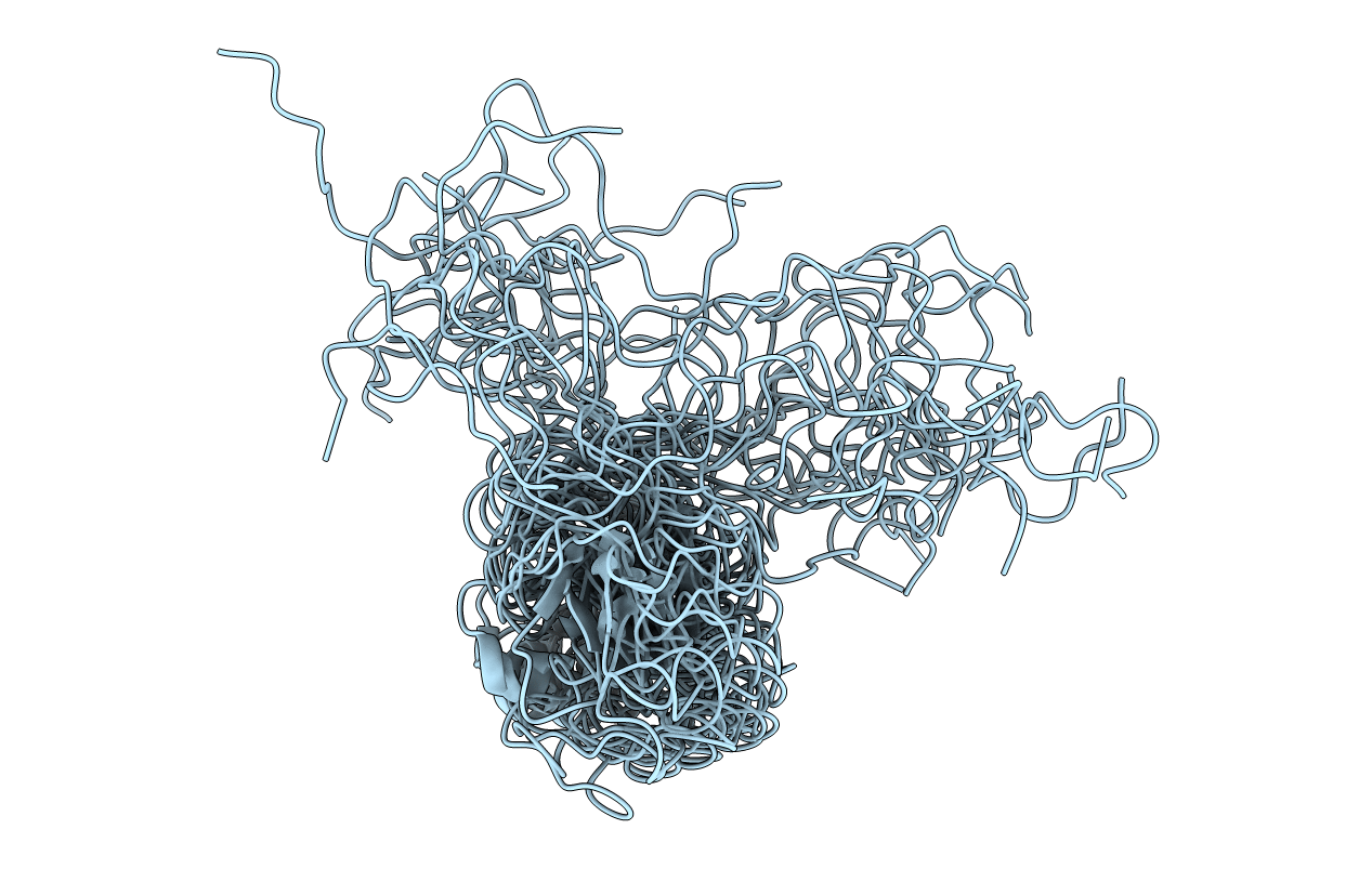
PDB ID:
1JFN
Keywords:
Title:
SOLUTION STRUCTURE OF HUMAN APOLIPOPROTEIN(A) KRINGLE IV TYPE 6
Biological Source:
Source Organism(s):
Homo sapiens (Taxon ID: 9606)
Expression System(s):
Method Details:
Experimental Method:
Conformers Calculated:
150
Conformers Submitted:
15
Selection Criteria:
structures with the lowest energy