Deposition Date
2001-06-20
Release Date
2001-09-19
Last Version Date
2023-08-16
Entry Detail
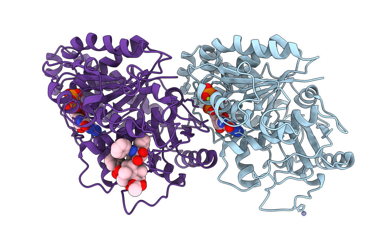
PDB ID:
1JFF
Keywords:
Title:
Refined structure of alpha-beta tubulin from zinc-induced sheets stabilized with taxol
Biological Source:
Source Organism(s):
Bos taurus (Taxon ID: 9913)
Method Details:
Experimental Method:
Resolution:
3.50 Å
R-Value Free:
0.29
R-Value Work:
0.23
Space Group:
P 1 21 1