1JCZ image
Deposition Date 2001-06-11
Release Date 2001-08-17
Last Version Date 2024-11-20
Entry Detail
PDB ID:
1JCZ
Keywords:
Title:
CRYSTAL STRUCTURE OF THE EXTRACELLULAR DOMAIN OF HUMAN CARBONIC ANHYDRASE XII
Biological Source:
Source Organism:
Homo sapiens (Taxon ID: 9606)
Method Details:
Experimental Method:
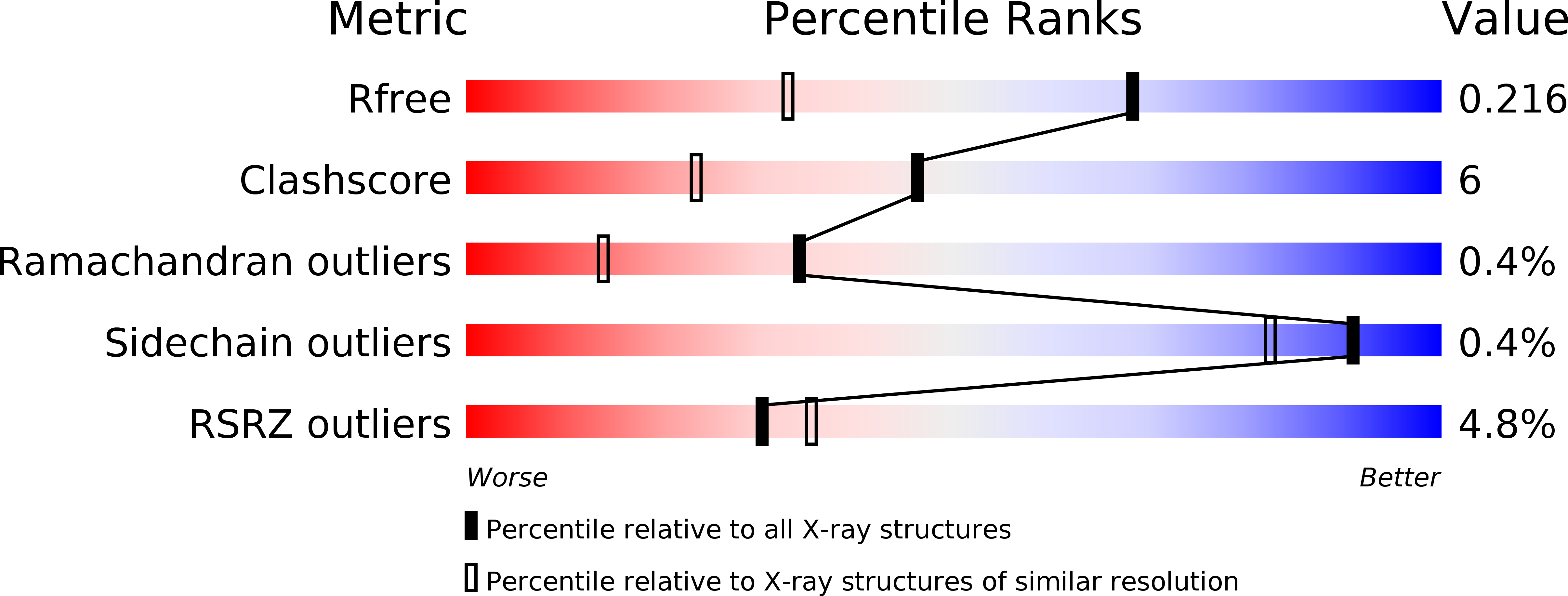
Resolution:
1.55 Å
R-Value Free:
0.22
R-Value Work:
0.19
R-Value Observed:
0.19
Space Group:
C 1 2 1
Macromolecular Entities
Polymer Type:polypeptide(L)
Molecule:CARBONIC ANHYDRASE XII
Gene (Uniprot):CA12
Chain IDs:A, B
Chain Length:263
Number of Molecules:2
Biological Source:Homo sapiens
Primary Citation
Crystal structure of the dimeric extracellular domain of human carbonic anhydrase XII, a bitopic membrane protein overexpressed in certain cancer tumor cells.
Proc.Natl.Acad.Sci.USA 98 9545 9550 (2001)
PMID: 11493685 DOI: 10.1073/pnas.161301298

Abstact

Overexpression of the zinc enzyme carbonic anhydrase (CA; EC ) XII is observed in certain human cancers. This bitopic membrane protein contains an N-terminal extracellular catalytic domain, a membrane-spanning alpha-helix, and a small intracellular C-terminal domain. We have determined the three-dimensional structure of the extracellular catalytic domain of human CA XII by x-ray crystallographic methods at 1.55-A resolution. The structure reveals a prototypical CA fold; however, two CA XII domains associate to form an isologous dimer, an observation that is confirmed by studies of the enzyme in solution. The identification of signature GXXXG and GXXXS motifs in the transmembrane sequence that facilitate helix-helix association is additionally consistent with dimeric architecture. The dimer interface is situated so that the active site clefts of each monomer are clearly exposed on one face of the dimer, and the C termini are located together on the opposite face of the dimer to facilitate membrane interaction. The amino acid composition of the active-site cleft closely resembles that of the other CA isozymes in the immediate vicinity of the catalytic zinc ion, but differs in the region of the nearby alpha-helical "130's segment." The structure of the CA XII-acetazolamide complex is also reported at 1.50-A resolution, and prospects for the design of CA XII-specific inhibitors of possible chemotherapeutic value are discussed.

Legend

Protein

Chemical

Disease

Primary Citation of related structures