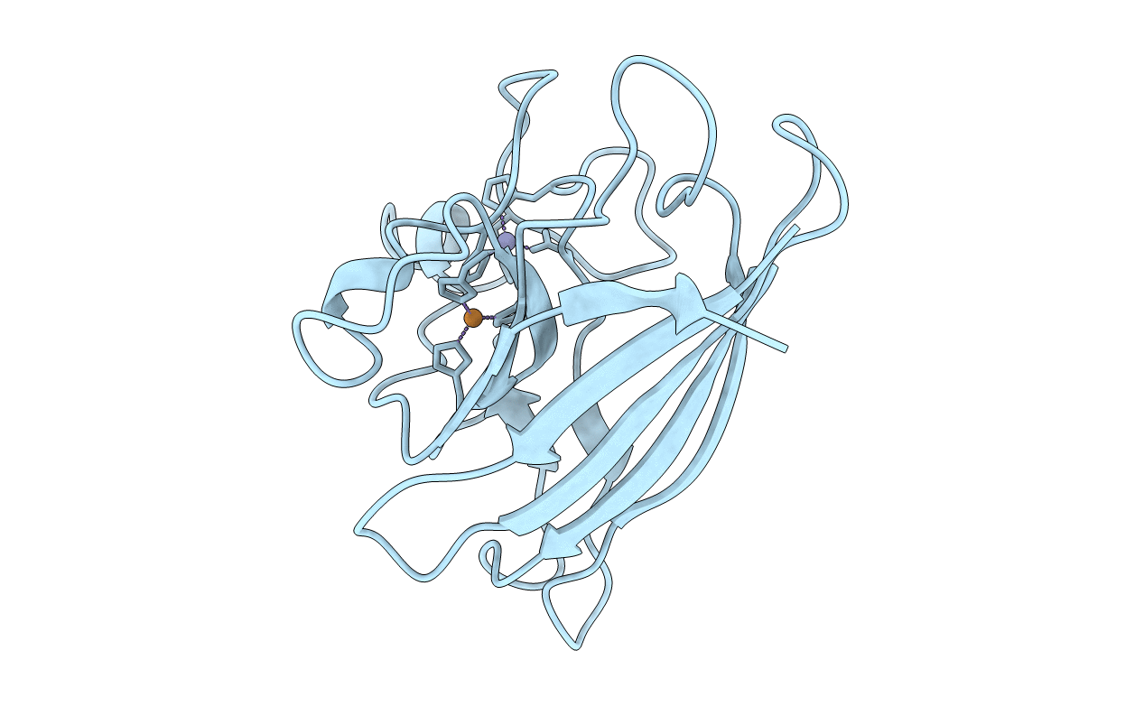
1JCV image
Entry Detail
PDB ID:
1JCV
Title:
REDUCED BRIDGE-BROKEN YEAST CU/ZN SUPEROXIDE DISMUTASE LOW TEMPERATURE (-180C) STRUCTURE
Biological Source:
Source Organism:
PDB Version:
Deposition Date:
1995-12-07
Release Date:
1996-03-08
Method Details:
Experimental Method:
Resolution:
1.55 Å
Space Group:
H 3 2
Macromolecular Entities
Polymer Type:polypeptide(L)
Description:CU/ZN SUPEROXIDE DISMUTASE
Chain IDs:A
Chain Length:153
Number of Molecules:1
Biological Source:Saccharomyces cerevisiae
Primary Citation
Unusual trigonal-planar copper configuration revealed in the atomic structure of yeast copper-zinc superoxide dismutase.
Biochemistry 35 2316 2321 (1996)
PMID: 8652572 DOI: 10.1021/bi951930b

Abstact

The three-dimensional structure of yeast copper-zinc superoxide dismutase (CuZnSOD) has been determined in a new crystal form in space group R32 and refined against X-ray diffraction data using difference Fourier and restrained crystallographic refinement techniques. The unexpected result is that the copper ion has moved approximately 1 angstrom from its position in previously reported CuZnSOD models, the copper-imidazolate bridge is broken, and a roughly trigonal planar ligand geometry characteristic of Cu(I) rather than Cu(II) is revealed. Final R values for the two nearly identical room temperature structures are 18.6% for all 19 149 reflections in the 10.0-1.7 angstrom resolution range and 18. 2% for 17 682 reflections (F > 2 sigma) in the 10.0-1.73 angstrom resolution range. A third structure has been determined using X-ray data collected at -180 degrees C. The final R value for this structure is 19.0% (R(free) = 22.9%) for all 24 356 reflections in the 10.0-1.55 angstrom resolution range. Virtually no change in the positions of the ligands to the zinc center is observed in these models. The origin of the broken bridge and altered Cu-ligand geometry is discussed.

Legend

Protein

Chemical

Disease

Primary Citation of related structures