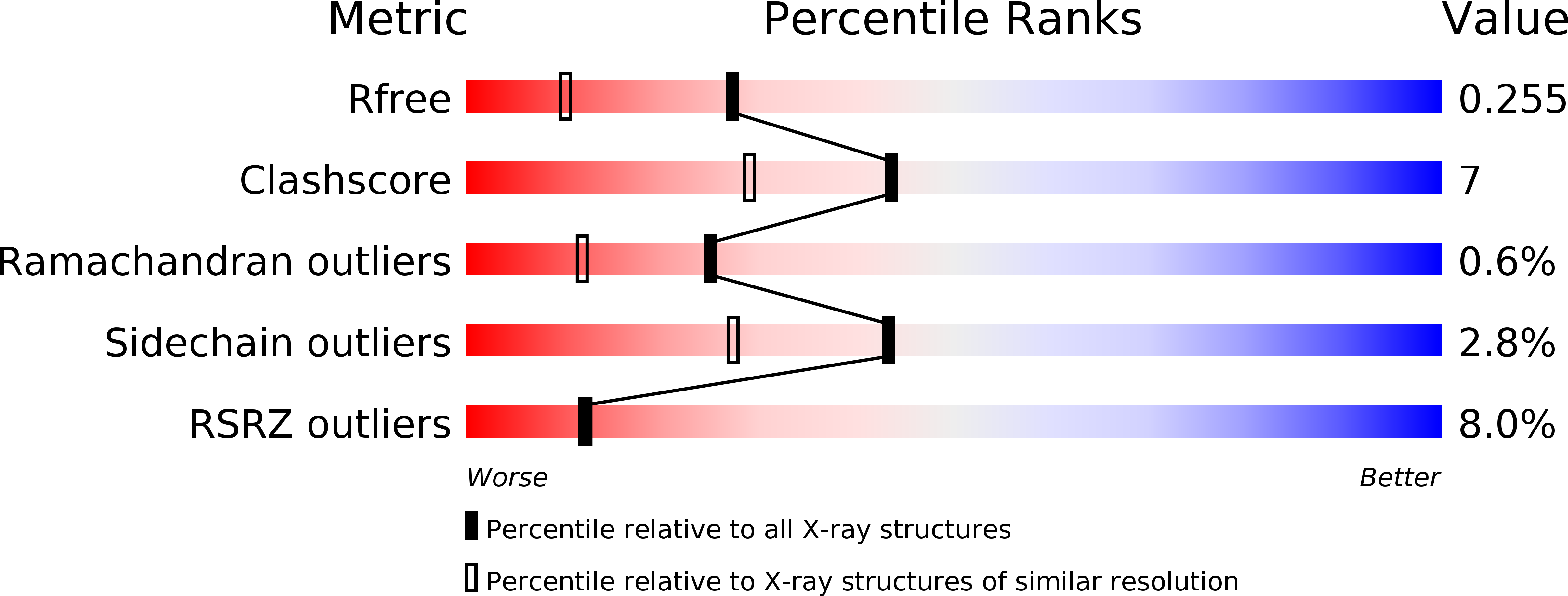
1JB7 image
Deposition Date 2001-06-02
Release Date 2001-06-11
Last Version Date 2023-08-16
Entry Detail
PDB ID:
1JB7
Title:
DNA G-Quartets in a 1.86 A Resolution Structure of an Oxytricha nova Telomeric Protein-DNA Complex
Biological Source:
Source Organism:
Sterkiella nova (Taxon ID: 200597)
(Taxon ID: )
Host Organism:
Method Details:
Experimental Method:
Resolution:
1.86 Å
R-Value Free:
0.24
R-Value Work:
0.23
R-Value Observed:
0.23
Space Group:
P 61 2 2
Macromolecular Entities
Polymer Type:polypeptide(L)
Molecule:telomere-binding protein alpha subunit
Gene (Uniprot):MAC-56A, MAC-56K, MAC-56S
Chain IDs:D (auth: A)
Chain Length:495
Number of Molecules:1
Biological Source:Sterkiella nova
Polymer Type:polypeptide(L)
Molecule:telomere-binding protein beta subunit
Gene (Uniprot):MAC-41A, MAC-41S
Chain IDs:E (auth: B)
Chain Length:260
Number of Molecules:1
Biological Source:Sterkiella nova
Polymer Type:polydeoxyribonucleotide
Molecule:5'-D(*GP*GP*GP*GP*TP*TP*TP*TP*GP*GP*GP*G)-3'
Chain IDs:A (auth: D), B (auth: G), C (auth: H)
Chain Length:12
Number of Molecules:3
Biological Source:
Primary Citation
DNA G-quartets in a 1.86 A resolution structure of an Oxytricha nova telomeric protein-DNA complex.
J.Mol.Biol. 310 367 377 (2001)
PMID: 11428895 DOI: 10.1006/jmbi.2001.4766

Abstact

The Oxytricha nova telomere end binding protein (OnTEBP) recognizes, binds and protects the single-stranded 3'-terminal DNA extension found at the ends of macronuclear chromosomes. The structure of this complex shows that the single strand GGGGTTTTGGGG DNA binds in a deep cleft between the two protein subunits of OnTEBP, adopting a non-helical and irregular conformation. In extending the resolution limit of this structure to 1.86 A, we were surprised to find a G-quartet linked dimer of the GGGGTTTTGGGG DNA also packing within the crystal lattice and interacting with the telomere end binding protein. The G-quartet DNA exhibits the same structure and topology as previously observed in solution by NMR with diagonally crossing d(TTTT) loops at either end of the four-stranded helix. Additionally, the crystal structure reveals clearly visible Na(+), and specific patterns of bound water molecules in the four non-equivalent grooves. Although the G-quartet:protein contact surfaces are modest and might simply represent crystal packing interactions, it is interesting to speculate that the two types of telomeric DNA-protein interactions observed here might both be important in telomere biology.

Legend

Protein

Chemical

Disease

Primary Citation of related structures