1JB6 image
Entry Detail
PDB ID:
1JB6
Keywords:
Title:
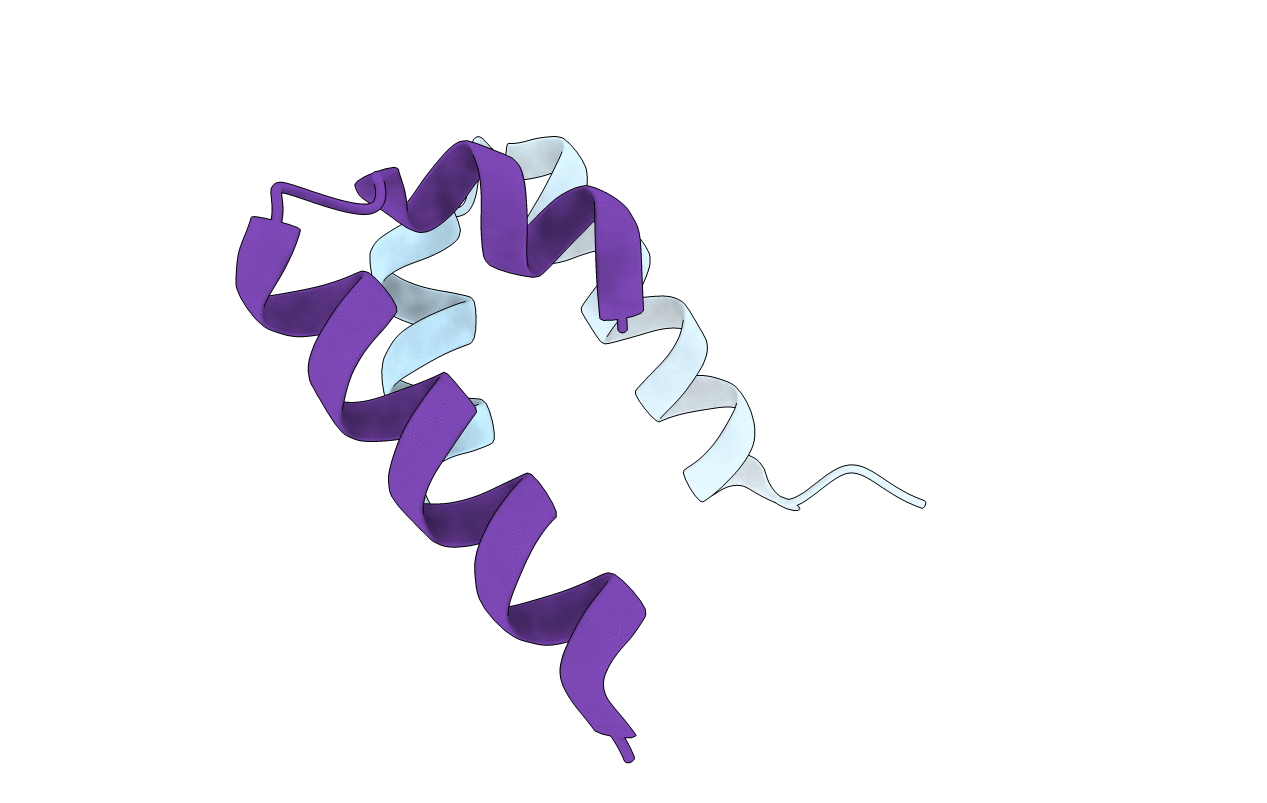
Crystal Structure of Dimerization Domain (1-33) of HNF-1alpha
Biological Source:
Source Organism:
PDB Version:
Deposition Date:
2001-06-01
Release Date:
2001-07-11
Method Details:
Experimental Method:
Resolution:
1.70 Å
R-Value Free:
0.24
R-Value Work:
0.23
Space Group:
P 21 21 2
Macromolecular Entities
Polymer Type:polypeptide(L)
Description:HEPATOCYTE NUCLEAR FACTOR 1-ALPHA
Chain IDs:A, B
Chain Length:33
Number of Molecules:2
Biological Source:
Modified Residue
Compound ID Chain ID Parent Comp ID Details 2D Image
MSE A MET SELENOMETHIONINE
NLE A LEU NORLEUCINE
Primary Citation
The dimerization domain of HNF-1alpha: structure and plasticity of an intertwined four-helix bundle with application to diabetes mellitus.
J.Mol.Biol. 310 635 658 (2001)
PMID: 11439029 DOI: 10.1006/jmbi.2001.4780

Abstact

Maturity-onset diabetes mellitus of the young (MODY) is a human genetic syndrome most commonly due to mutations in hepatocyte nuclear factor-1alpha (HNF-1alpha). Here, we describe the crystal structure of the HNF-1alpha dimerization domain at 1.7 A resolution and assess its structural plasticity. The crystal's low solvent content (23%, v/v) leads to tight packing of peptides in the lattice. Two independent dimers, similar in structure, are formed in the unit cell by a 2-fold crystallographic symmetry axis. The dimers define a novel intertwined four-helix bundle (4HB). Each protomer contains two alpha-helices separated by a sharp non-canonical turn. Dimer-related alpha-helices form anti-parallel coiled-coils, including an N-terminal "mini-zipper" complementary in structure, symmetry and surface characteristics to transcriptional coactivator dimerization cofactor of HNF-1 (DCoH). A confluence of ten leucine side-chains (five per protomer) forms a hydrophobic core. Isotope-assisted NMR studies demonstrate that a similar intertwined dimer exists in solution. Comparison of structures obtained in multiple independent crystal forms indicates that the mini-zipper is a stable structural element, whereas the C-terminal alpha-helix can adopt a broad range of orientations. Segmental alignment of the mini-zipper (mean pairwise root-mean-square difference (rmsd) in C(alpha) coordinates of 0.29 A) is associated with a 2.1 A mean C(alpha) rmsd displacement of the C-terminal coiled-coil. The greatest C-terminal structural variation (4.1 A C(alpha) rmsd displacement) is observed in the DCoH-bound peptide. Diabetes-associated mutations perturb distinct structural features of the HNF-1alpha domain. One mutation (L12H) destabilizes the domain but preserves structural specificity. Adjoining H12 side-chains in a native-like dimer are predicted to alter the functional surface of the mini-zipper involved in DCoH recognition. The other mutation (G20R), by contrast, leads to a dimeric molten globule, as indicated by its 1H-NMR features and fluorescent binding of 1-anilino-8-naphthalene sulfonate. We propose that a glycine-specific turn configuration enables specific interactions between the mini-zipper and the C-terminal coiled-coil.

Legend

Protein

Chemical

Disease

Primary Citation of related structures