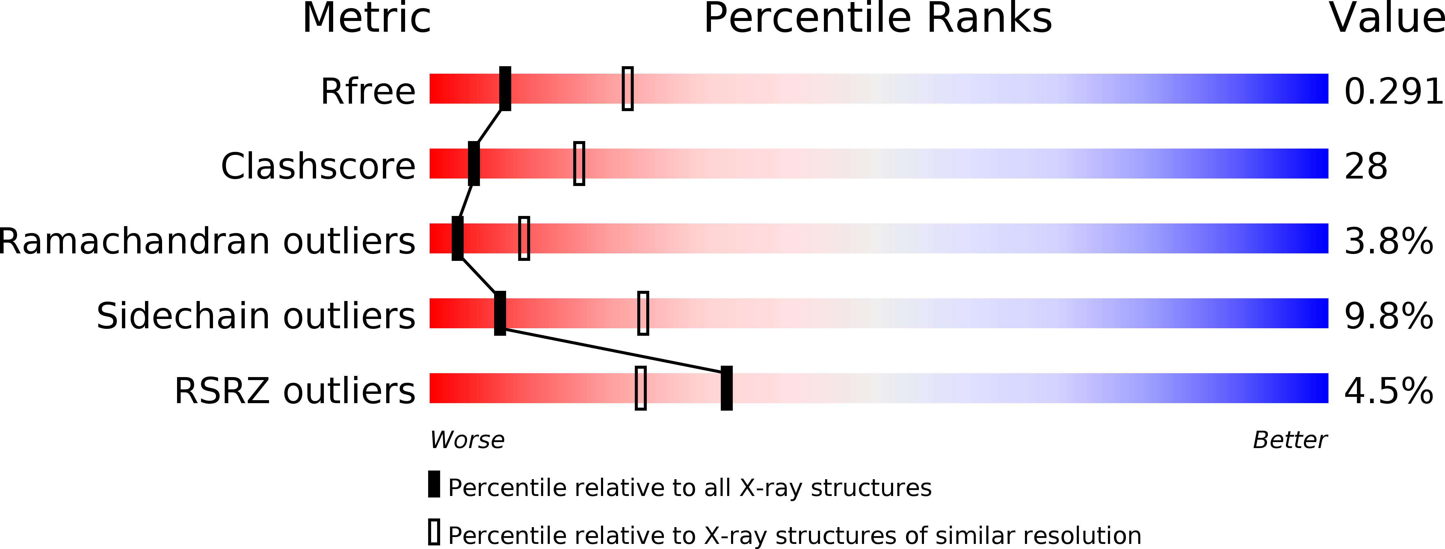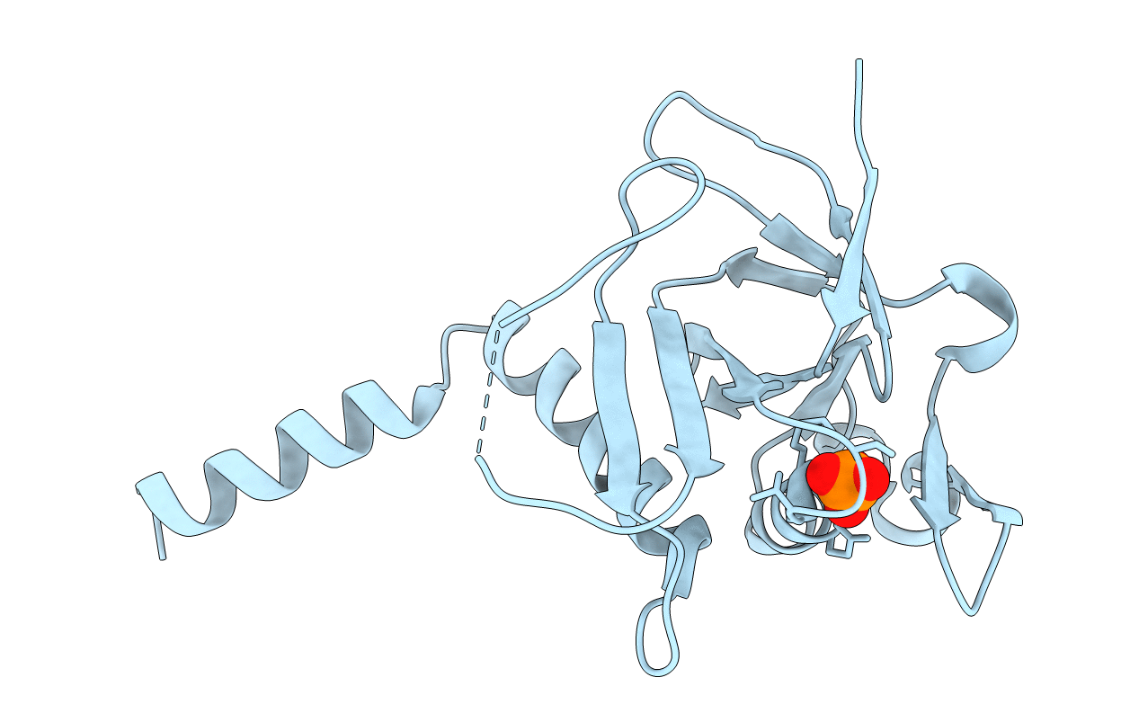
1JB1 image
Deposition Date 2001-06-01
Release Date 2001-08-08
Last Version Date 2024-11-06
Entry Detail
PDB ID:
1JB1
Title:
Lactobacillus casei HprK/P Bound to Phosphate
Biological Source:
Source Organism:
Host Organism:
Method Details:
Experimental Method:
Resolution:
2.80 Å
R-Value Free:
0.28
R-Value Work:
0.23
Space Group:
P 63 2 2
Macromolecular Entities
Polymer Type:polypeptide(L)
Molecule:HPRK PROTEIN
Gene (Uniprot):hprK
Chain IDs:A
Chain Length:205
Number of Molecules:1
Biological Source:Lactobacillus casei
Primary Citation
X-ray structure of HPr kinase: a bacterial protein kinase with a P-loop nucleotide-binding domain.
EMBO J. 20 3917 3927 (2001)
PMID: 11483495 DOI: 10.1093/emboj/20.15.3917

Abstact

HPr kinase/phosphatase (HprK/P) is a key regulatory enzyme controlling carbon metabolism in Gram- positive bacteria. It catalyses the ATP-dependent phosphorylation of Ser46 in HPr, a protein of the phosphotransferase system, and also its dephosphorylation. HprK/P is unrelated to eukaryotic protein kinases, but contains the Walker motif A characteristic of nucleotide-binding proteins. We report here the X-ray structure of an active fragment of Lactobacillus casei HprK/P at 2.8 A resolution, solved by the multiwavelength anomalous dispersion method on a seleniated protein (PDB code 1jb1). The protein is a hexamer, with each subunit containing an ATP-binding domain similar to nucleoside/nucleotide kinases, and a putative HPr-binding domain unrelated to the substrate-binding domains of other kinases. The Walker motif A forms a typical P-loop which binds inorganic phosphate in the crystal. We modelled ATP binding by comparison with adenylate kinase, and designed a tentative model of the complex with HPr based on a docking simulation. The results confirm that HprK/P represents a new family of protein kinases, first identified in bacteria, but which may also have members in eukaryotes.

Legend

Protein

Chemical

Disease

Primary Citation of related structures