Deposition Date
2001-06-01
Release Date
2003-09-09
Last Version Date
2024-11-13
Entry Detail
PDB ID:
1JAZ
Keywords:
Title:
Crystal Structure of Monoclinic Form of D90E Mutant of Escherichia coli Asparaginase II
Biological Source:
Source Organism:
Escherichia coli (Taxon ID: 562)
Host Organism:
Method Details:
Experimental Method:
Resolution:
2.27 Å
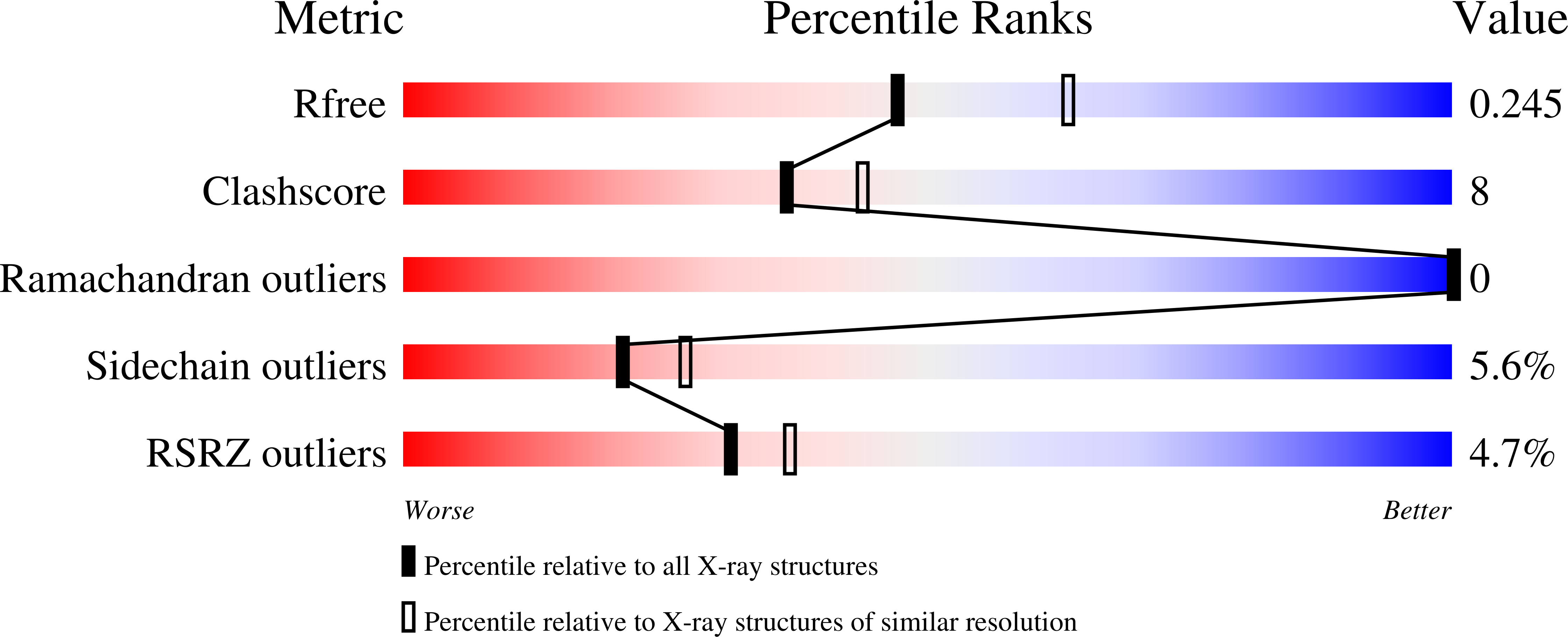
R-Value Free:
0.23
R-Value Work:
0.18
R-Value Observed:
0.18
Space Group:
C 1 2 1