1JAP image
Deposition Date 1996-03-11
Release Date 1996-07-11
Last Version Date 2024-06-05
Entry Detail
PDB ID:
1JAP
Title:
COMPLEX OF PRO-LEU-GLY-HYDROXYLAMINE WITH THE CATALYTIC DOMAIN OF MATRIX METALLO PROTEINASE-8 (MET80 FORM)
Biological Source:
Source Organism:
Homo sapiens (Taxon ID: 9606)
Host Organism:
Method Details:
Experimental Method:
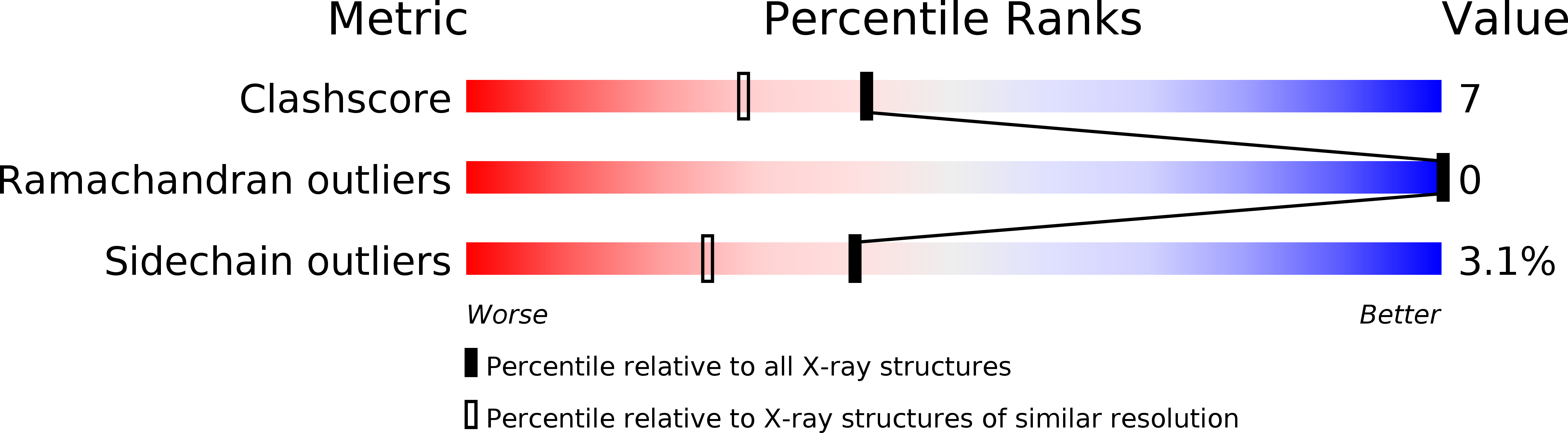
Resolution:
1.82 Å
R-Value Work:
0.19
R-Value Observed:
0.19
Space Group:
P 21 21 21
Macromolecular Entities
Polymer Type:polypeptide(L)
Molecule:MATRIX METALLO PROTEINASE-8 (MET80 FORM)
Gene (Uniprot):MMP8
Chain IDs:A
Chain Length:163
Number of Molecules:1
Biological Source:Homo sapiens
Peptide-like Molecules
PRD_000404
Primary Citation
The X-ray crystal structure of the catalytic domain of human neutrophil collagenase inhibited by a substrate analogue reveals the essentials for catalysis and specificity.
EMBO J. 13 1263 1269 (1994)
PMID: 8137810

Abstact

Matrix metalloproteinases are a family of zinc endopeptidases involved in tissue remodelling. They have been implicated in various disease processes including tumour invasion and joint destruction. These enzymes consist of several domains, which are responsible for latency, catalysis and substrate recognition. Human neutrophil collagenase (PMNL-CL, MMP-8) represents one of the two 'interstitial' collagenases that cleave triple helical collagens types I, II and III. Its 163 residue catalytic domain (Met80 to Gly242) has been expressed in Escherichia coli and crystallized as a non-covalent complex with the inhibitor Pro-Leu-Gly-hydroxylamine. The 2.0 A crystal structure reveals a spherical molecule with a shallow active-site cleft separating a smaller C-terminal subdomain from a bigger N-terminal domain, composed of a five-stranded beta-sheet, two alpha-helices, and bridging loops. The inhibitor mimics the unprimed (P1-P3) residues of a substrate; primed (P1'-P3') peptide substrate residues should bind in an extended conformation, with the bulky P1' side-chain fitting into the deep hydrophobic S1' subsite. Modelling experiments with collagen show that the scissile strand of triple-helical collagen must be freed to fit the subsites. The catalytic zinc ion is situated at the bottom of the active-site cleft and is penta-coordinated by three histidines and by both hydroxamic acid oxygens of the inhibitor. In addition to the catalytic zinc, the catalytic domain harbours a second, non-exchangeable zinc ion and two calcium ions, which are packed against the top of the beta-sheet and presumably function to stabilize the catalytic domain.(ABSTRACT TRUNCATED AT 250 WORDS)

Legend

Protein

Chemical

Disease

Primary Citation of related structures