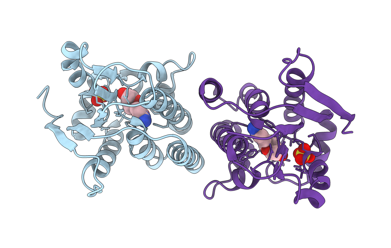
1J90 image
Entry Detail
PDB ID:
1J90
Keywords:
Title:
Crystal Structure of Drosophila Deoxyribonucleoside Kinase
Biological Source:
Source Organism:
Host Organism:
PDB Version:
Deposition Date:
2001-05-23
Release Date:
2001-11-28
Method Details:
Experimental Method:
Resolution:
2.56 Å
R-Value Free:
0.25
R-Value Work:
0.23
R-Value Observed:
0.26
Space Group:
P 21 21 2
Macromolecular Entities
Polymer Type:polypeptide(L)
Description:Deoxyribonucleoside kinase
Mutations:Deletion of last 20 residues
Chain IDs:A, B
Chain Length:230
Number of Molecules:2
Biological Source:Drosophila melanogaster
Primary Citation
Structural basis for substrate specificities of cellular deoxyribonucleoside kinases.
Nat.Struct.Biol. 8 616 620 (2001)
PMID: 11427893 DOI: 10.1038/89661

Abstact

Deoxyribonucleoside kinases phosphorylate deoxyribonucleosides and activate a number of medically important nucleoside analogs. Here we report the structure of the Drosophila deoxyribonucleoside kinase with deoxycytidine bound at the nucleoside binding site and that of the human deoxyguanosine kinase with ATP at the nucleoside substrate binding site. Compared to the human kinase, the Drosophila kinase has a wider substrate cleft, which may be responsible for the broad substrate specificity of this enzyme. The human deoxyguanosine kinase is highly specific for purine substrates; this is apparently due to the presence of Arg 118, which provides favorable hydrogen bonding interactions with the substrate. The two new structures provide an explanation for the substrate specificity of cellular deoxyribonucleoside kinases.

Legend

Protein

Chemical

Disease

Primary Citation of related structures