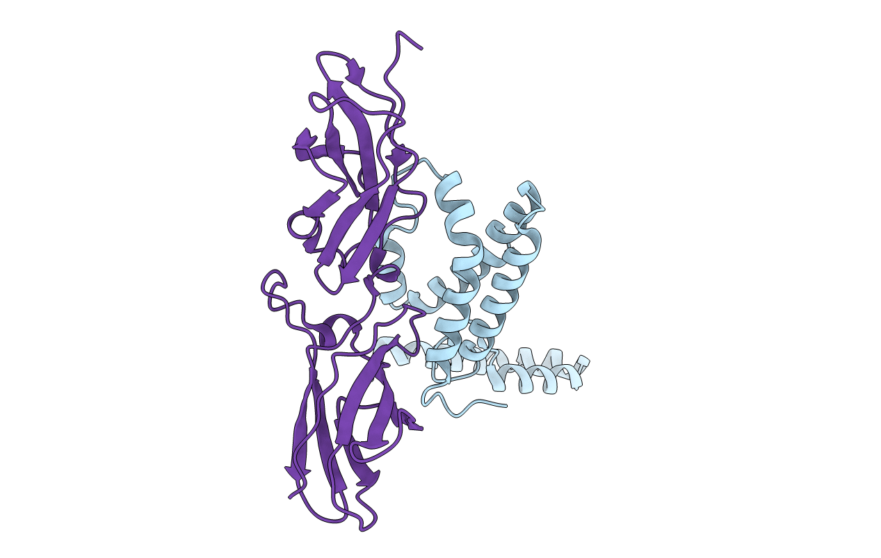
1J7V image
Deposition Date 2001-05-18
Release Date 2001-09-19
Last Version Date 2024-10-30
Entry Detail
PDB ID:
1J7V
Title:
HUMAN IL-10 / IL-10R1 COMPLEX
Biological Source:
Source Organism:
Homo sapiens (Taxon ID: 9606)
Host Organism:
Method Details:
Experimental Method:
Resolution:
2.90 Å
R-Value Free:
0.28
R-Value Work:
0.23
Space Group:
P 32 1 2
Macromolecular Entities
Polymer Type:polypeptide(L)
Molecule:INTERLEUKIN-10
Gene (Uniprot):IL10
Chain IDs:A (auth: L)
Chain Length:160
Number of Molecules:1
Biological Source:Homo sapiens
Polymer Type:polypeptide(L)
Molecule:INTERLEUKIN-10 RECEPTOR ALPHA CHAIN
Gene (Uniprot):IL10RA
Mutations:N29Q,N53Q,N89Q,N133Q,N156Q,N168Q
Chain IDs:B (auth: R)
Chain Length:214
Number of Molecules:1
Biological Source:Homo sapiens
Primary Citation
Crystal structure of the IL-10/IL-10R1 complex reveals a shared receptor binding site.
Immunity 15 35 46 (2001)
PMID: 11485736 DOI: 10.1016/S1074-7613(01)00169-8

Abstact

Interleukin 10 (IL-10) is a dimeric cytokine that plays a central role in suppressing inflammatory responses. These activities are dependent on the interaction of IL-10 with its high-affinity receptor (IL-10R1). This intermediate complex must subsequently recruit the low-affinity IL-10R2 chain before cell signaling can occur. Here we report the 2.9 A crystal structure of IL-10 bound to a soluble form of IL-10R1 (sIL-10R1). The complex consists of two IL-10s and four sIL-10R1 molecules. Several residues in the IL-10/sIL-10R1 interface are conserved in all IL-10 homologs and their receptors. The data suggests that formation of the active IL-10 signaling complex occurs by a novel molecular recognition paradigm where IL-10R1 and IL-10R2 both recognize the same binding site on IL-10.

Legend

Protein

Chemical

Disease

Primary Citation of related structures