1J79 image
Deposition Date 2001-05-16
Release Date 2001-06-20
Last Version Date 2025-03-26
Entry Detail
PDB ID:
1J79
Keywords:
Title:
Molecular Structure of Dihydroorotase: A Paradigm for Catalysis Through the Use of a Binuclear Metal Center
Biological Source:
Source Organism:
Escherichia coli (Taxon ID: 562)
Host Organism:
Method Details:
Experimental Method:
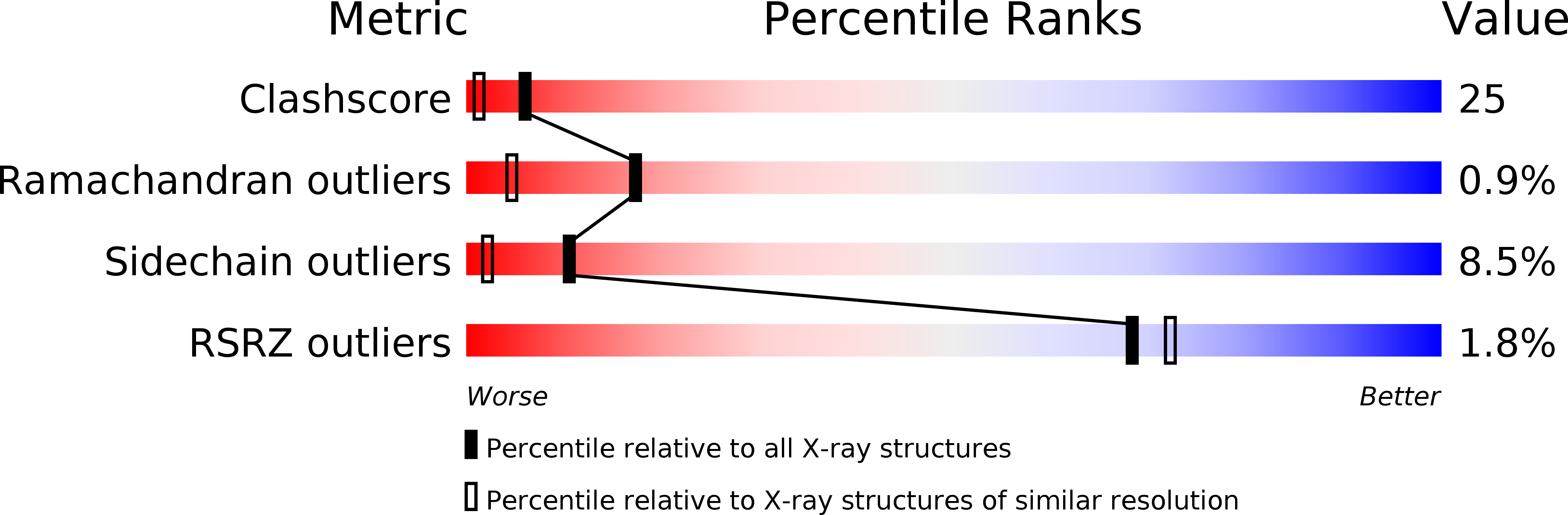
Resolution:
1.70 Å
R-Value Free:
0.25
R-Value Work:
0.19
R-Value Observed:
0.19
Space Group:
P 21 21 21
Macromolecular Entities
Structures with similar UniProt ID
Protein Blast
Polymer Type:polypeptide(L)
Molecule:dihydroorotase
Gene (Uniprot):pyrC
Chain IDs:A, B
Chain Length:347
Number of Molecules:2
Biological Source:Escherichia coli
Modified Residue
Compound ID Chain ID Parent Comp ID Details 2D Image
KCX A LYS LYSINE NZ-CARBOXYLIC ACID
Primary Citation
Molecular structure of dihydroorotase: a paradigm for catalysis through the use of a binuclear metal center.
Biochemistry 40 6989 6997 (2001)
PMID: 11401542 DOI: 10.1021/bi010682i

Abstact

Dihydroorotase plays a key role in pyrimidine biosynthesis by catalyzing the reversible interconversion of carbamoyl aspartate to dihydroorotate. Here we describe the three-dimensional structure of dihydroorotase from Escherichia coli determined and refined to 1.7 A resolution. Each subunit of the homodimeric enzyme folds into a "TIM" barrel motif with eight strands of parallel beta-sheet flanked on the outer surface by alpha-helices. Unexpectedly, each subunit contains a binuclear zinc center with the metal ions separated by approximately 3.6 A. Lys 102, which is carboxylated, serves as a bridging ligand between the two cations. The more buried or alpha-metal ion in subunit I is surrounded by His 16, His 18, Lys 102, Asp 250, and a solvent molecule (most likely a hydroxide ion) in a trigonal bipyramidal arrangement. The beta-metal ion, which is closer to the solvent, is tetrahedrally ligated by Lys 102, His 139, His 177, and the bridging hydroxide. L-Dihydroorotate is observed bound to subunit I, with its carbonyl oxygen, O4, lying 2.9 A from the beta-metal ion. Important interactions for positioning dihydroorotate into the active site include a salt bridge with the guanidinium group of Arg 20 and various additional electrostatic interactions with both protein backbone and side chain atoms. Strikingly, in subunit II, carbamoyl L-aspartate is observed binding near the binuclear metal center with its carboxylate side chain ligating the two metals and thus displacing the bridging hydroxide ion. From the three-dimensional structures of the enzyme-bound substrate and product, it has been possible to propose a unique catalytic mechanism for dihydroorotase. In the direction of dihydroorotate hydrolysis, the bridging hydroxide attacks the re-face of dihydroorotate with general base assistance by Asp 250. The carbonyl group is polarized for nucleophilic attack by the bridging hydroxide through a direct interaction with the beta-metal ion. During the cyclization of carbamoyl aspartate, Asp 250 initiates the reaction by abstracting a proton from N3 of the substrate. The side chain carboxylate of carbamoyl aspartate is polarized through a direct electrostatic interaction with the binuclear metal center. The ensuing tetrahedral intermediate collapses with C-O bond cleavage and expulsion of the hydroxide which then bridges the binuclear metal center.

Legend

Protein

Chemical

Disease

Primary Citation of related structures