1J77 image
Deposition Date 2001-05-15
Release Date 2001-05-30
Last Version Date 2024-02-07
Entry Detail
PDB ID:
1J77
Keywords:
Title:
Crystal Structure of Gram-negative Bacterial Heme Oxygenase Complexed with Heme
Biological Source:
Source Organism:
Method Details:
Experimental Method:
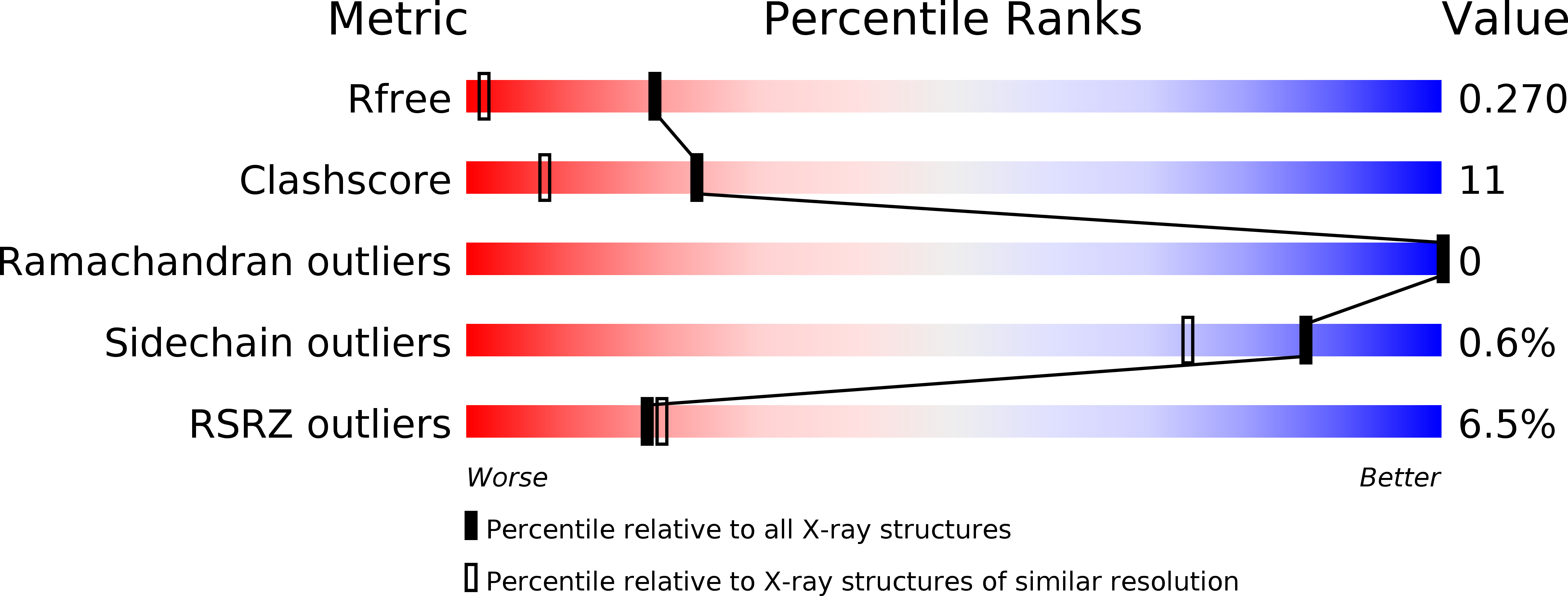
Resolution:
1.50 Å
R-Value Free:
0.26
R-Value Work:
0.22
R-Value Observed:
0.22
Space Group:
P 43 21 2
Macromolecular Entities
Polymer Type:polypeptide(L)
Molecule:HemO
Gene (Uniprot):hemO
Chain IDs:A
Chain Length:209
Number of Molecules:1
Biological Source:Neisseria meningitidis
Ligand Molecules
Primary Citation
Crystal structure of heme oxygenase from the gram-negative pathogen Neisseria meningitidis and a comparison with mammalian heme oxygenase-1.
Biochemistry 40 11552 11558 (2001)
PMID: 11560504 DOI: 10.1021/bi0110239

Abstact

We report the crystal structure of heme oxygenase from the pathogenic bacterium Neisseria meningitidis at 1.5 A and compare and contrast it with known structures of heme oxygenase-1 from mammalian sources. Both the bacterial and mammalian enzymes share the same overall fold, with a histidine contributing a ligand to the proximal side of the heme iron and a kinked alpha-helix defining the distal pocket. The distal helix differs noticeably in both sequence and conformation, and the distal pocket of the Neisseria enzyme is substantially smaller than in the mammalian enzyme. Key glycine residues provide the flexibility for the helical kink, allow close contact of the helix backbone with the heme, and may interact directly with heme ligands.

Legend

Protein

Chemical

Disease

Primary Citation of related structures