Deposition Date
2001-05-15
Release Date
2001-09-01
Last Version Date
2023-08-16
Entry Detail
PDB ID:
1J75
Keywords:
Title:
Crystal Structure of the DNA-Binding Domain Zalpha of DLM-1 Bound to Z-DNA
Biological Source:
Source Organism(s):
Mus musculus (Taxon ID: 10090)
Expression System(s):
Method Details:
Experimental Method:
Resolution:
1.85 Å
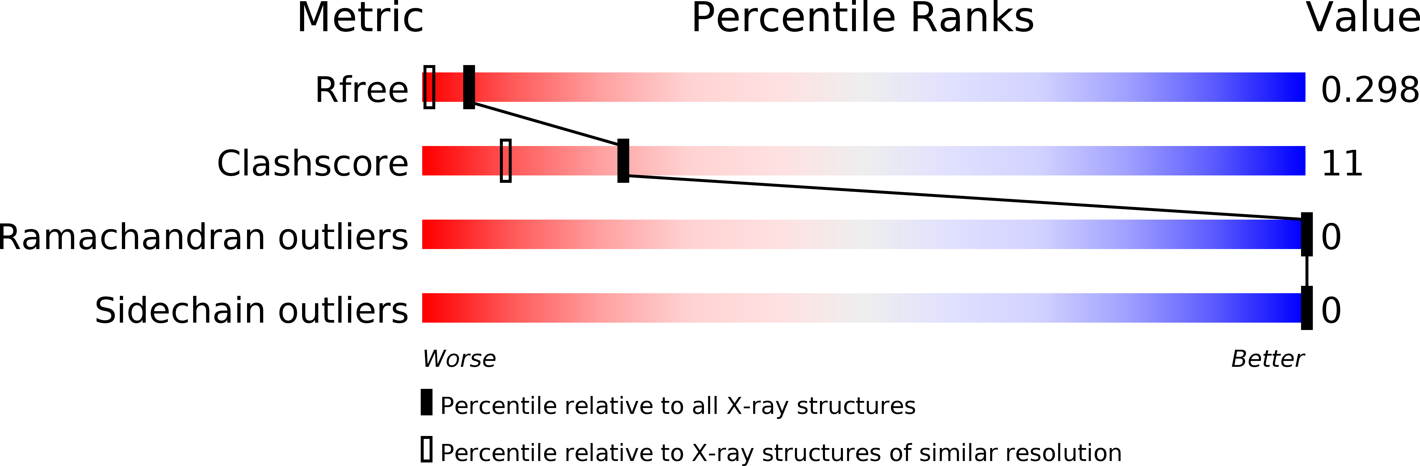
R-Value Free:
0.24
R-Value Work:
0.21
R-Value Observed:
0.22
Space Group:
P 65 2 2