1J71 image
Deposition Date 2001-05-15
Release Date 2001-05-23
Last Version Date 2024-10-16
Entry Detail
PDB ID:
1J71
Keywords:
Title:
Structure of the extracellular aspartic proteinase from Candida tropicalis yeast.
Biological Source:
Source Organism:
Candida tropicalis (Taxon ID: 5482)
unidentified (Taxon ID: 32644)
Method Details:
Experimental Method:
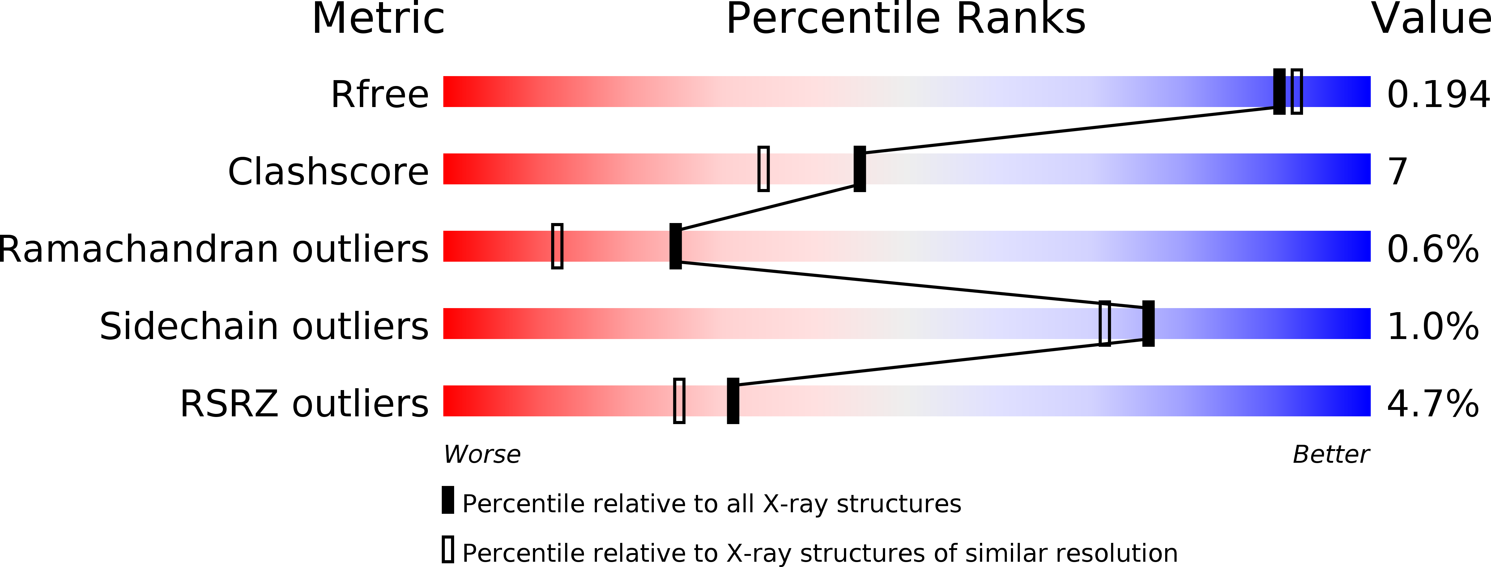
Resolution:
1.80 Å
R-Value Free:
0.19
R-Value Work:
0.16
R-Value Observed:
0.16
Space Group:
P 21 21 21
Macromolecular Entities
Polymer Type:polypeptide(L)
Molecule:Aspartic proteinase
Gene (Uniprot):SAPT1
Chain IDs:A
Chain Length:334
Number of Molecules:1
Biological Source:Candida tropicalis
Polymer Type:polypeptide(L)
Molecule:Tetrapeptide Thr-Ile-Thr-Ser
Chain IDs:B
Chain Length:4
Number of Molecules:1
Biological Source:unidentified
Ligand Molecules
Primary Citation
High-resolution structure of the extracellular aspartic proteinase from Candida tropicalis yeast.
Biochemistry 36 12700 12710 (1997)
PMID: 9335526 DOI: 10.1021/bi970613x

Abstact

The crystal structure of the secreted aspartic proteinase from Candida tropicalis yeast (SAPT) has been determined to 1.8 A resolution. The classic aspartic proteinase bilobal structure and domain topology is conserved in SAPT, with the substrate binding cleft situated between the two domains. Structural comparisons made with pepsin indicate that insertions and deletions in the primary sequence modify the SAPT structure to create a more spacious substrate binding cleft with altered specificity. An unexpected tetrapeptide has been found to occupy binding sites S1'-S3', and this suggests the order of release of peptide products in the catalytic mechanism of these enzymes. Structural features are considered with regard to previous substrate specificity data.

Legend

Protein

Chemical

Disease

Primary Citation of related structures