
Deposition Date
2003-01-31
Release Date
2003-02-18
Last Version Date
2023-12-27
Entry Detail
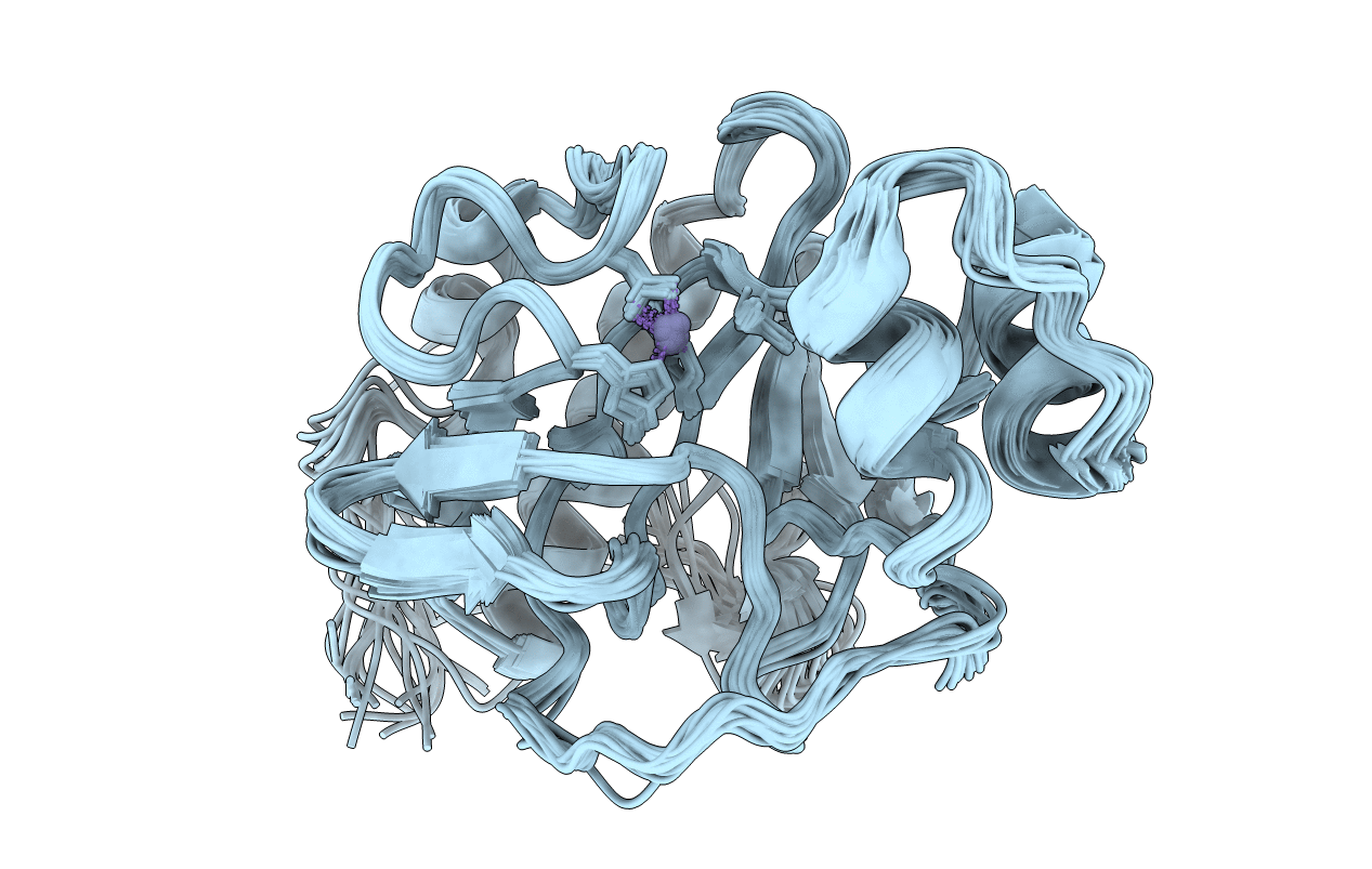
Biological Source:
Source Organism(s):
Citrobacter freundii (Taxon ID: 546)
Expression System(s):
Method Details:
Experimental Method:
Conformers Calculated:
20
Conformers Submitted:
20
Selection Criteria:
all calculated structures submitted
