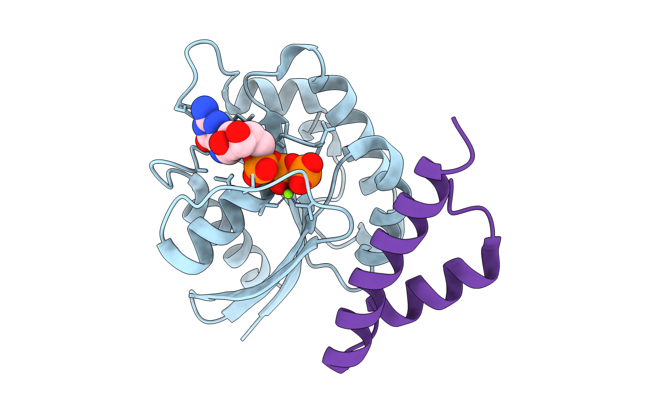
1J2J image
Entry Detail
PDB ID:
1J2J
Title:
Crystal structure of GGA1 GAT N-terminal region in complex with ARF1 GTP form
Biological Source:
Source Organism:
Host Organism:
PDB Version:
Deposition Date:
2003-01-05
Release Date:
2003-05-06
Method Details:
Experimental Method:
Resolution:
1.60 Å
R-Value Free:
0.22
R-Value Work:
0.19
R-Value Observed:
0.19
Space Group:
P 21 21 21
Macromolecular Entities
Polymer Type:polypeptide(L)
Description:ADP-ribosylation factor 1
Mutations:Q71L
Chain IDs:A
Chain Length:166
Number of Molecules:1
Biological Source:Mus musculus
Polymer Type:polypeptide(L)
Description:ADP-ribosylation factor binding protein GGA1
Chain IDs:B
Chain Length:45
Number of Molecules:1
Biological Source:Homo sapiens
Primary Citation
Molecular mechanism of membrane recruitment of GGA by ARF in lysosomal protein transport
NAT.STRUCT.BIOL. 10 386 393 (2003)
PMID: 12679809 DOI: 10.1038/nsb920

Abstact

GGAs are critical for trafficking soluble proteins from the trans-Golgi network (TGN) to endosomes/lysosomes through interactions with TGN-sorting receptors, ADP-ribosylation factor (ARF) and clathrin. ARF-GTP bound to TGN membranes recruits its effector GGA by binding to the GAT domain, thus facilitating recognition of GGA for cargo-loaded receptors. Here we report the X-ray crystal structures of the human GGA1-GAT domain and the complex between ARF1-GTP and the N-terminal region of the GAT domain. When unbound, the GAT domain forms an elongated bundle of three a-helices with a hydrophobic core. Structurally, this domain, combined with the preceding VHS domain, resembles CALM, an AP180 homolog involved in endocytosis. In the complex with ARF1-GTP, a helix-loop-helix of the N-terminal part of GGA1-GAT interacts with the switches 1 and 2 of ARF1 predominantly in a hydrophobic manner. These data reveal a molecular mechanism underlying membrane recruitment of adaptor proteins by ARF-GTP.

Legend

Protein

Chemical

Disease

Primary Citation of related structures