1J2C image
Deposition Date 2002-12-29
Release Date 2003-09-02
Last Version Date 2023-10-25
Entry Detail
PDB ID:
1J2C
Keywords:
Title:
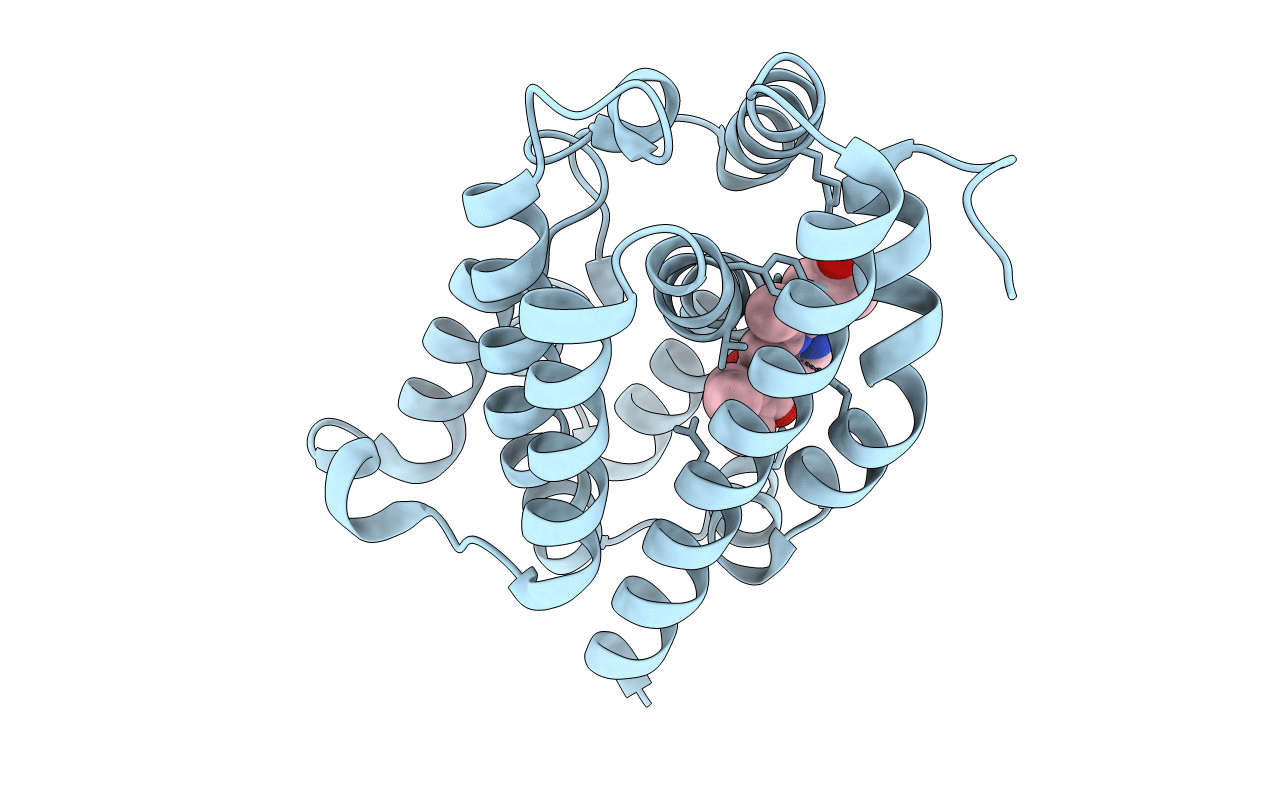
Crystal structure of rat heme oxygenase-1 in complex with biliverdin IXalpha-iron cluster
Biological Source:
Source Organism:
Host Organism:
Method Details:
Experimental Method:
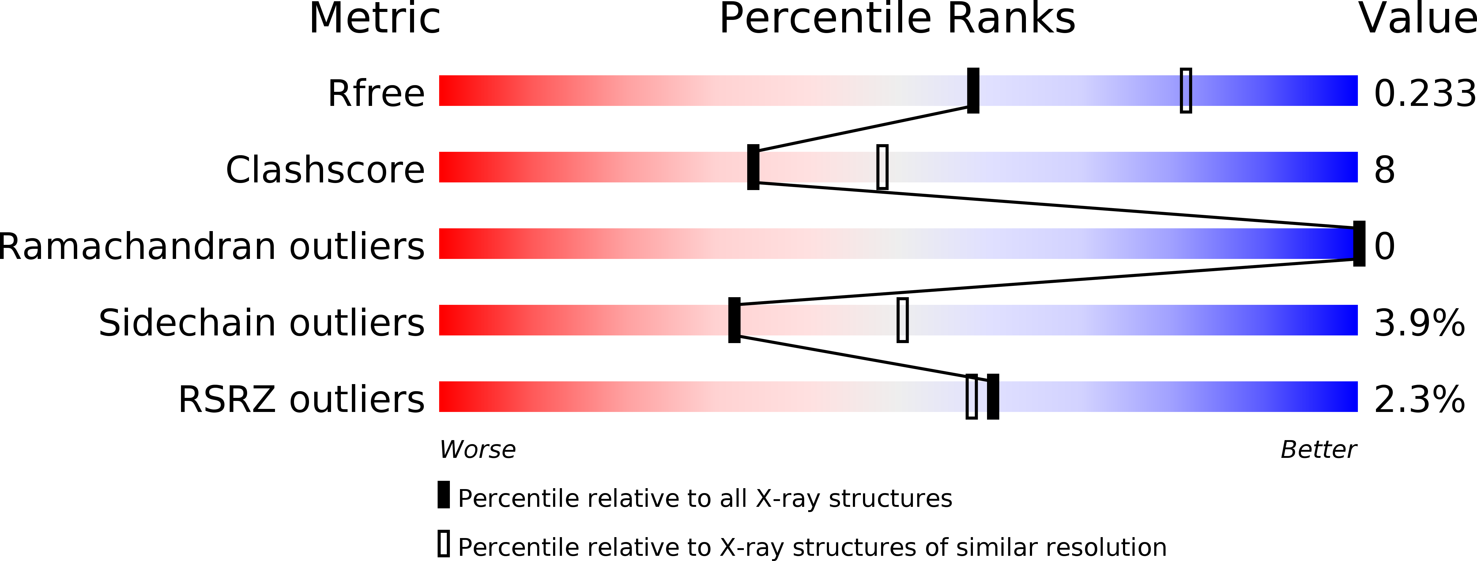
Resolution:
2.40 Å
R-Value Free:
0.23
R-Value Work:
0.19
R-Value Observed:
0.19
Space Group:
P 32 2 1
Macromolecular Entities
Polymer Type:polypeptide(L)
Molecule:Heme Oxygenase-1
Gene (Uniprot):Hmox1
Chain IDs:A
Chain Length:267
Number of Molecules:1
Biological Source:Rattus norvegicus
Primary Citation
Crystal Structure of Rat Heme Oxygenase-1 in Complex with Biliverdin-Iron Chelate: CONFORMATIONAL CHANGE OF THE DISTAL HELIX DURING THE HEME CLEAVAGE REACTION.
J.Biol.Chem. 278 32352 32358 (2003)
PMID: 12794075 DOI: 10.1074/jbc.M303682200

Abstact

The crystal structure of rat heme oxygenase-1 in complex with biliverdin-iron chelate (biliverdin(Fe)-HO-1), the immediate precursor of the final product, biliverdin, has been determined at a 2.4-A resolution. The electron density in the heme pocket clearly showed that the tetrapyrrole ring of heme is cleaved at the alpha-meso edge. Like the heme bound to HO-1, biliverdin-iron chelate is located between the distal and proximal helices, but its accommodation state seems to be less stable in light of the disordering of the solvent-exposed propionate and vinyl groups. The middle of the distal helix is shifted away from the center of the active site in biliverdin(Fe)-HO-1, increasing the size of the heme pocket. The hydrogen-bonding interaction between Glu-29 and Gln-38, considered to restrain the orientation of the proximal helix in the heme-HO-1 complex, was lost in biliverdin(Fe)-HO-1, leading to relaxation of the helix. Biliverdin has a distorted helical conformation; the lactam oxygen atom of its pyrrole ring-A interacted with Asp-140 through a hydrogen-bonding solvent network. Because of the absence of a distal water ligand, the iron atom is five-coordinated with His-25 and four pyrrole nitrogen atoms. The coordination geometry deviates considerably from a square pyramid, suggesting that the iron may be readily dissociated. We speculate that the opened conformation of the heme pocket facilitates sequential product release, first iron then biliverdin, and that because of biliverdin's increased flexibility, iron release triggers its slow dissociation.

Legend

Protein

Chemical

Disease

Primary Citation of related structures