1J1V image
Deposition Date 2002-12-18
Release Date 2003-04-22
Last Version Date 2024-10-30
Entry Detail
PDB ID:
1J1V
Keywords:
Title:
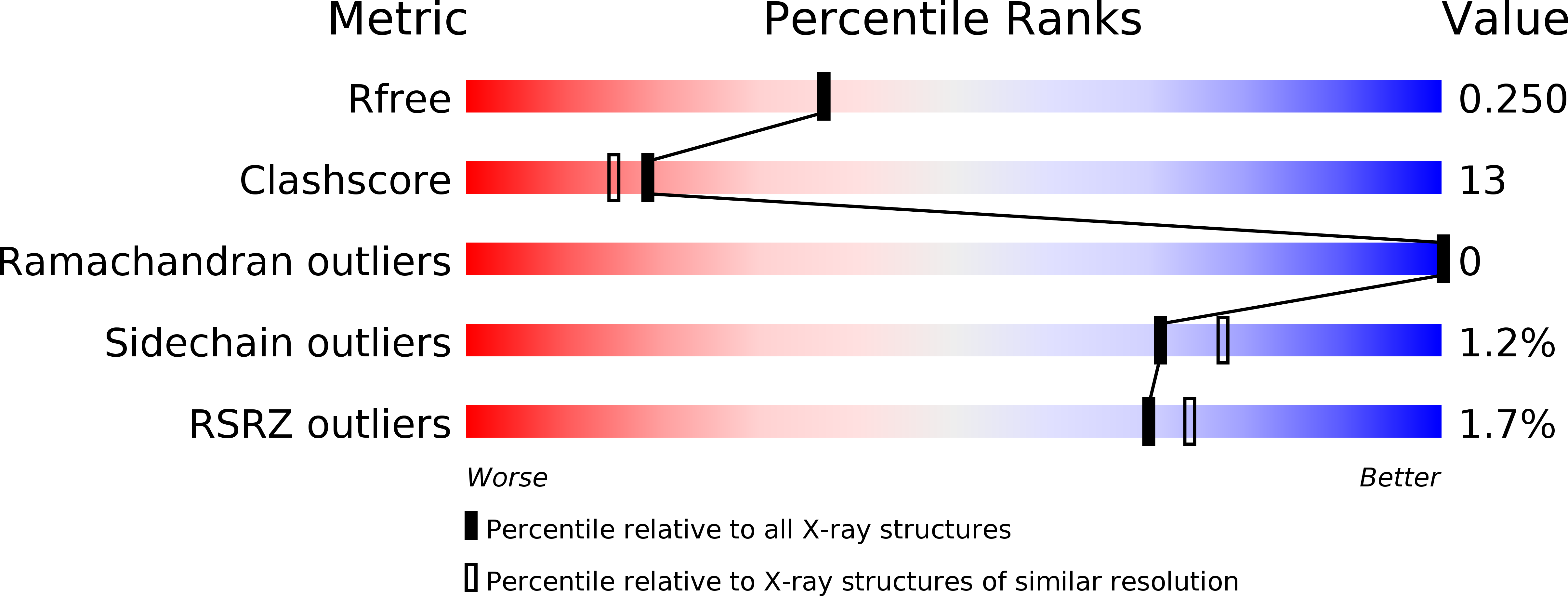
Crystal structure of DnaA domainIV complexed with DnaAbox DNA
Biological Source:
Source Organism:
Escherichia coli (Taxon ID: 562)
Method Details:
Experimental Method:
Resolution:
2.10 Å
R-Value Free:
0.24
R-Value Work:
0.22
R-Value Observed:
0.22
Space Group:
P 41 21 2
Macromolecular Entities
Polymer Type:polypeptide(L)
Molecule:Chromosomal replication initiator protein dnaA
Gene (Uniprot):dnaA
Chain IDs:C (auth: A)
Chain Length:94
Number of Molecules:1
Biological Source:Escherichia coli
Modified Residue
Compound ID Chain ID Parent Comp ID Details 2D Image
MSE C MET SELENOMETHIONINE
Primary Citation
Structural basis of replication origin recognition by the DnaA protein
NUCLEIC ACIDS RES. 31 2077 2086 (2003)
PMID: 12682358 DOI: 10.1093/nar/gkg309

Abstact

Escherichia coli DnaA binds to 9 bp sequences (DnaA boxes) in the replication origin, oriC, to form a complex initiating chromosomal DNA replication. In the present study, we determined the crystal structure of its DNA-binding domain (domain IV) complexed with a DnaA box at 2.1 A resolution. DnaA domain IV contains a helix-turn-helix motif for DNA binding. One helix and a loop of the helix- turn-helix motif are inserted into the major groove and 5 bp (3' two-thirds of the DnaA box sequence) are recognized through base-specific hydrogen bonds and van der Waals contacts with the C5-methyl groups of thymines. In the minor groove, Arg399, located in the loop adjacent to the motif, recognizes three more base pairs (5' one-third of the DnaA box sequence) by base-specific hydrogen bonds. DNA bending by approximately 28 degrees was also observed in the complex. These base-specific interactions explain how DnaA exhibits higher affinity for the strong DnaA boxes (R1, R2 and R4) than the weak DnaA boxes (R3 and M) in the replication origin.

Legend

Protein

Chemical

Disease

Primary Citation of related structures