
Deposition Date
2002-11-25
Release Date
2003-06-17
Last Version Date
2024-11-06
Entry Detail
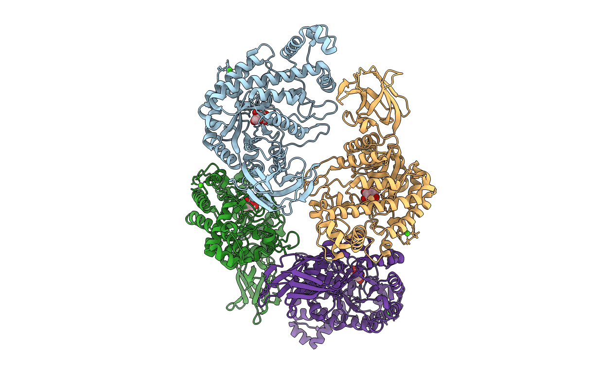
PDB ID:
1J0Y
Keywords:
Title:
Beta-amylase from Bacillus cereus var. mycoides in complex with glucose
Biological Source:
Source Organism(s):
Bacillus cereus (Taxon ID: 1396)
Method Details:
Experimental Method:
Resolution:
2.10 Å
R-Value Free:
0.23
R-Value Work:
0.17
Space Group:
C 1 2 1
