1IYI image
Entry Detail
PDB ID:
1IYI
Keywords:
Title:
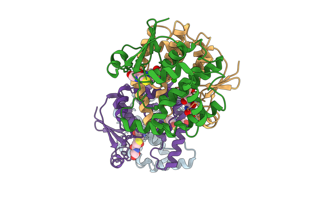
Crystal structure of hematopoietic prostaglandin D synthase
Biological Source:
Source Organism:
Host Organism:
PDB Version:
Deposition Date:
2002-08-26
Release Date:
2003-04-08
Method Details:
Experimental Method:
Resolution:
1.80 Å
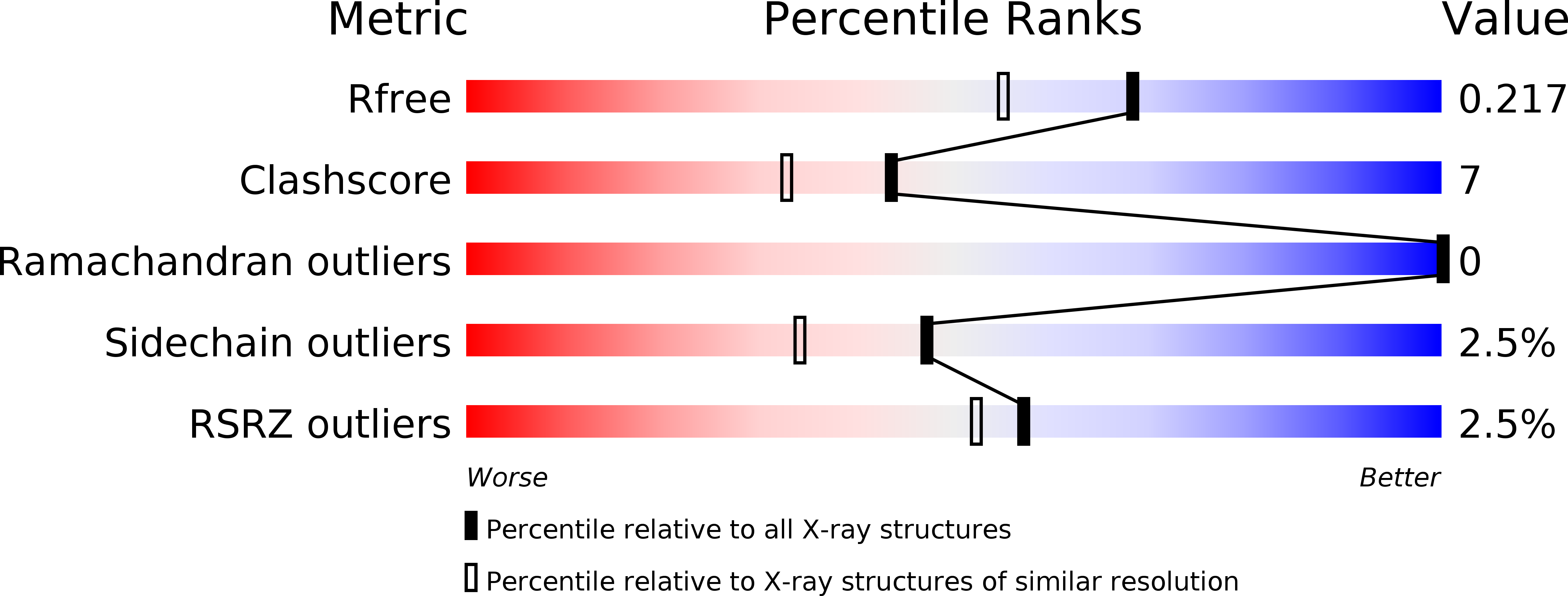
R-Value Free:
0.22
R-Value Work:
0.18
R-Value Observed:
0.18
Space Group:
P 1 21 1
Macromolecular Entities
Polymer Type:polypeptide(L)
Description:HEMATOPOIETIC PROSTAGLANDIN D SYNTHASE
Chain IDs:A, B, C, D
Chain Length:198
Number of Molecules:4
Biological Source:Homo sapiens
Primary Citation
Mechanism of metal activation of human hematopoietic prostaglandin D synthase
NAT.STRUCT.BIOL. 10 291 296 (2003)
PMID: 12627223 DOI: 10.1038/nsb907

Abstact

Here we report the crystal structures of human hematopoietic prostaglandin (PG) D synthase bound to glutathione (GSH) and Ca2+ or Mg2+. Using GSH as a cofactor, prostaglandin D synthase catalyzes the isomerization of PGH2 to PGD2, a mediator for allergy response. The enzyme is a homodimer, and Ca2+ or Mg2+ increases its activity to approximately 150% of the basal level, with half maximum effective concentrations of 400 microM for Ca2+ and 50 microM for Mg2+. In the Mg2+-bound form, the ion is octahedrally coordinated by six water molecules at the dimer interface. The water molecules are surrounded by pairs of Asp93, Asp96 and Asp97 from each subunit. Ca(2+) is coordinated by five water molecules and an Asp96 from one subunit. The Asp96 residue in the Ca2+-bound form makes hydrogen bonds with two guanidium nitrogen atoms of Arg14 in the GSH-binding pocket. Mg2+ alters the coordinating water structure and reduces one hydrogen bond between Asp96 and Arg14, thereby changing the interaction between Arg14 and GSH. This effect explains a four-fold reduction in the K(m) of the enzyme for GSH. The structure provides insights into how Ca2+ or Mg2+ binding activates human hematopoietic PGD synthase.

Legend

Protein

Chemical

Disease

Primary Citation of related structures