1IXS image
Entry Detail
PDB ID:
1IXS
Keywords:
Title:
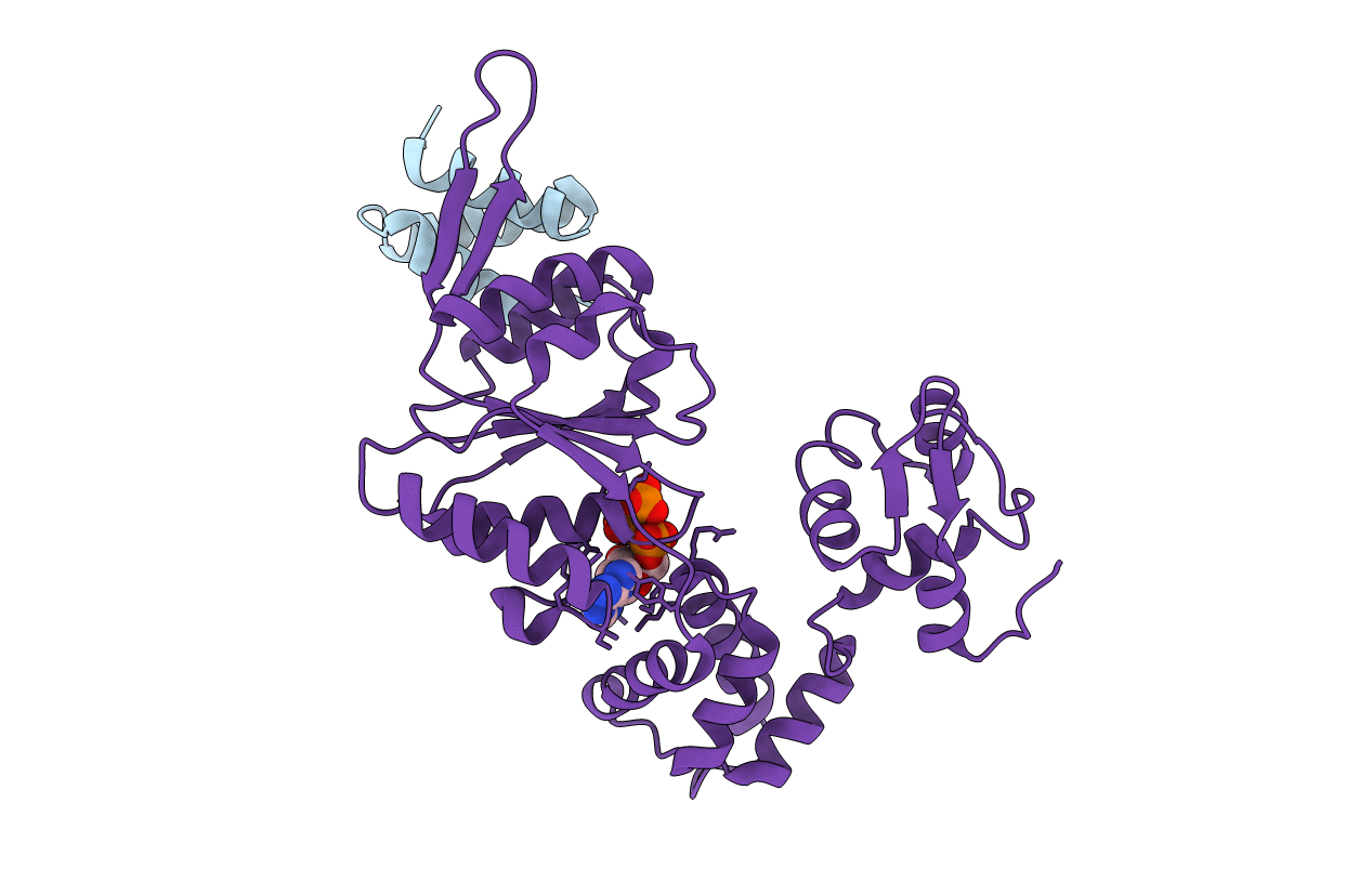
Structure of RuvB complexed with RuvA domain III
Biological Source:
Source Organism:
PDB Version:
Deposition Date:
2002-07-04
Release Date:
2002-11-06
Method Details:
Experimental Method:
Resolution:
3.20 Å
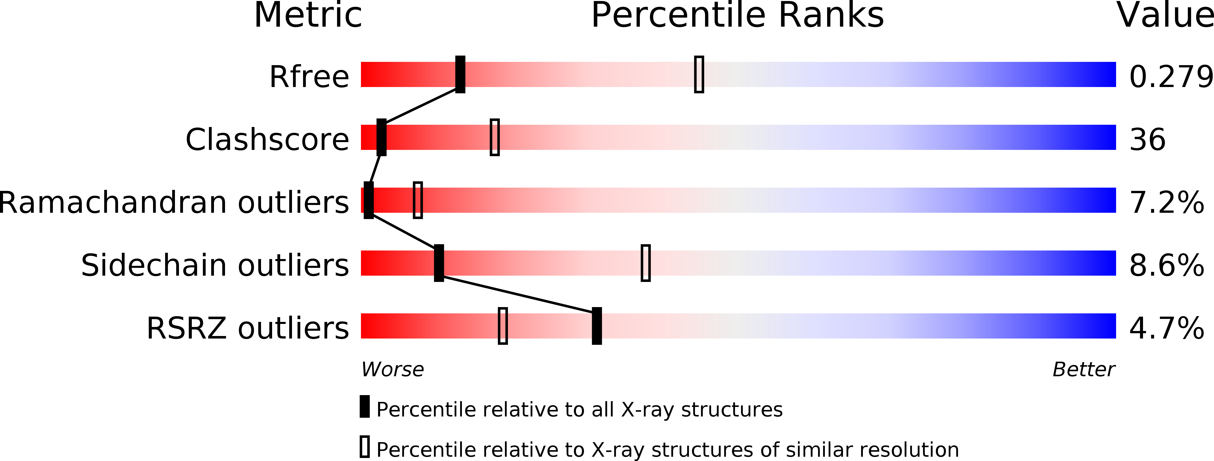
R-Value Free:
0.29
R-Value Work:
0.23
Space Group:
P 43 21 2
Macromolecular Entities
Polymer Type:polypeptide(L)
Description:Holliday junction DNA helicase ruvA
Chain IDs:A
Chain Length:62
Number of Molecules:1
Biological Source:Thermus thermophilus
Polymer Type:polypeptide(L)
Description:RuvB
Chain IDs:B
Chain Length:318
Number of Molecules:1
Biological Source:Thermus thermophilus
Ligand Molecules
Primary Citation
Crystal Structure of the RuvA-RuvB Complex: A Structural Basis for the Holliday Junction Migrating Motor Machinery
Mol.Cell 10 671 681 (2002)
PMID: 12408833 DOI: 10.1016/S1097-2765(02)00641-X

Abstact

We present the X-ray structure of the RuvA-RuvB complex, which plays a crucial role in ATP-dependent branch migration. Two RuvA tetramers form the symmetric and closed octameric shell, where four RuvA domain IIIs spring out in the two opposite directions to be individually caught by a single RuvB. The binding of domain III deforms the protruding beta hairpin in the N-terminal domain of RuvB and thereby appears to induce a functional and less symmetric RuvB hexameric ring. The model of the RuvA-RuvB junction DNA ternary complex, constructed by fitting the X-ray structure into the averaged electron microscopic images of the RuvA-RuvB junction, appears to be more compatible with the branch migration mode of a fixed RuvA-RuvB interaction than with a rotational interaction mode.

Legend

Protein

Chemical

Disease

Primary Citation of related structures