Deposition Date
2002-03-28
Release Date
2002-10-16
Last Version Date
2024-10-23
Entry Detail
PDB ID:
1IVO
Keywords:
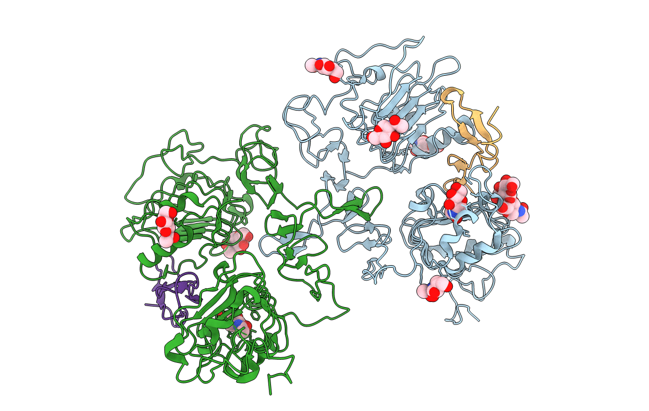
Title:
Crystal Structure of the Complex of Human Epidermal Growth Factor and Receptor Extracellular Domains.
Biological Source:
Source Organism(s):
Homo sapiens (Taxon ID: 9606)
Expression System(s):
Method Details:
Experimental Method:
Resolution:
3.30 Å
R-Value Free:
0.32
R-Value Work:
0.25
R-Value Observed:
0.25
Space Group:
P 31 2 1