Deposition Date
2002-03-01
Release Date
2003-09-30
Last Version Date
2023-12-27
Entry Detail
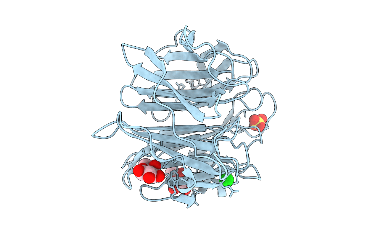
PDB ID:
1IUB
Keywords:
Title:
Fucose-specific lectin from Aleuria aurantia (Hg-derivative form)
Biological Source:
Source Organism(s):
Aleuria aurantia (Taxon ID: 5188)
Expression System(s):
Method Details:
Experimental Method:
Resolution:
2.31 Å
R-Value Free:
0.18
R-Value Work:
0.15
Space Group:
P 65 2 2