1ITU image
Entry Detail
PDB ID:
1ITU
Keywords:
Title:
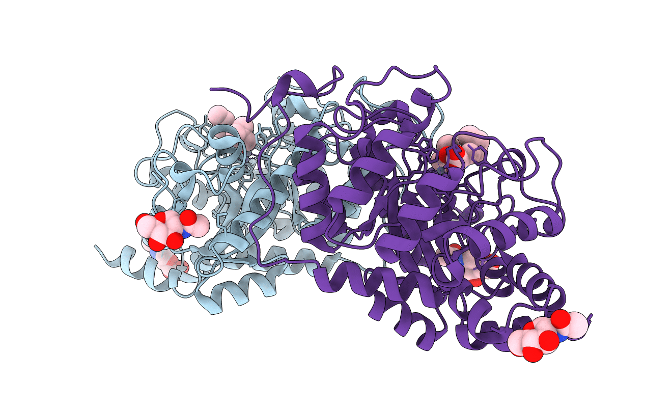
HUMAN RENAL DIPEPTIDASE COMPLEXED WITH CILASTATIN
Biological Source:
Source Organism:
Host Organism:
PDB Version:
Deposition Date:
2002-02-03
Release Date:
2002-08-28
Method Details:
Experimental Method:
Resolution:
2.00 Å
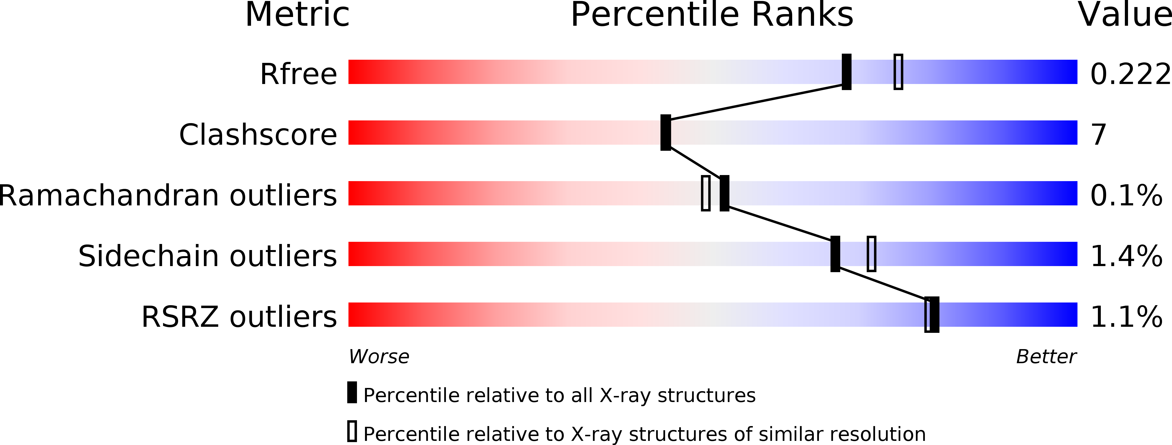
R-Value Free:
0.25
R-Value Work:
0.18
R-Value Observed:
0.18
Space Group:
P 1 21 1
Macromolecular Entities
Polymer Type:polypeptide(L)
Description:RENAL DIPEPTIDASE
Chain IDs:A, B
Chain Length:369
Number of Molecules:2
Biological Source:Homo sapiens
Modified Residue
Compound ID Chain ID Parent Comp ID Details 2D Image
ASN A ASN GLYCOSYLATION SITE
Primary Citation
Crystal Structure of Human Renal Dipeptidase Involved in beta-Lactam Hydrolysis
J.Mol.Biol. 321 177 184 (2002)
PMID: 12144777 DOI: 10.1016/S0022-2836(02)00632-0

Abstact

Human renal dipeptidase is a membrane-bound glycoprotein hydrolyzing dipeptides and is involved in hydrolytic metabolism of penem and carbapenem beta-lactam antibiotics. The crystal structures of the saccharide-trimmed enzyme are determined as unliganded and inhibitor-liganded forms. They are informative for designing new antibiotics that are not hydrolyzed by this enzyme. The active site in each of the (alpha/beta)(8) barrel subunits of the homodimeric molecule is composed of binuclear zinc ions bridged by the Glu125 side-chain located at the bottom of the barrel, and it faces toward the microvillar membrane of a kidney tubule. A dipeptidyl moiety of the therapeutically used cilastatin inhibitor is fully accommodated in the active-site pocket, which is small enough for precise recognition of dipeptide substrates. The barrel and active-site architectures utilizing catalytic metal ions exhibit unexpected similarities to those of the murine adenosine deaminase and the catalytic domain of the bacterial urease.

Legend

Protein

Chemical

Disease

Primary Citation of related structures