
Deposition Date
2002-01-09
Release Date
2002-05-22
Last Version Date
2023-10-25
Entry Detail
PDB ID:
1IT6
Keywords:
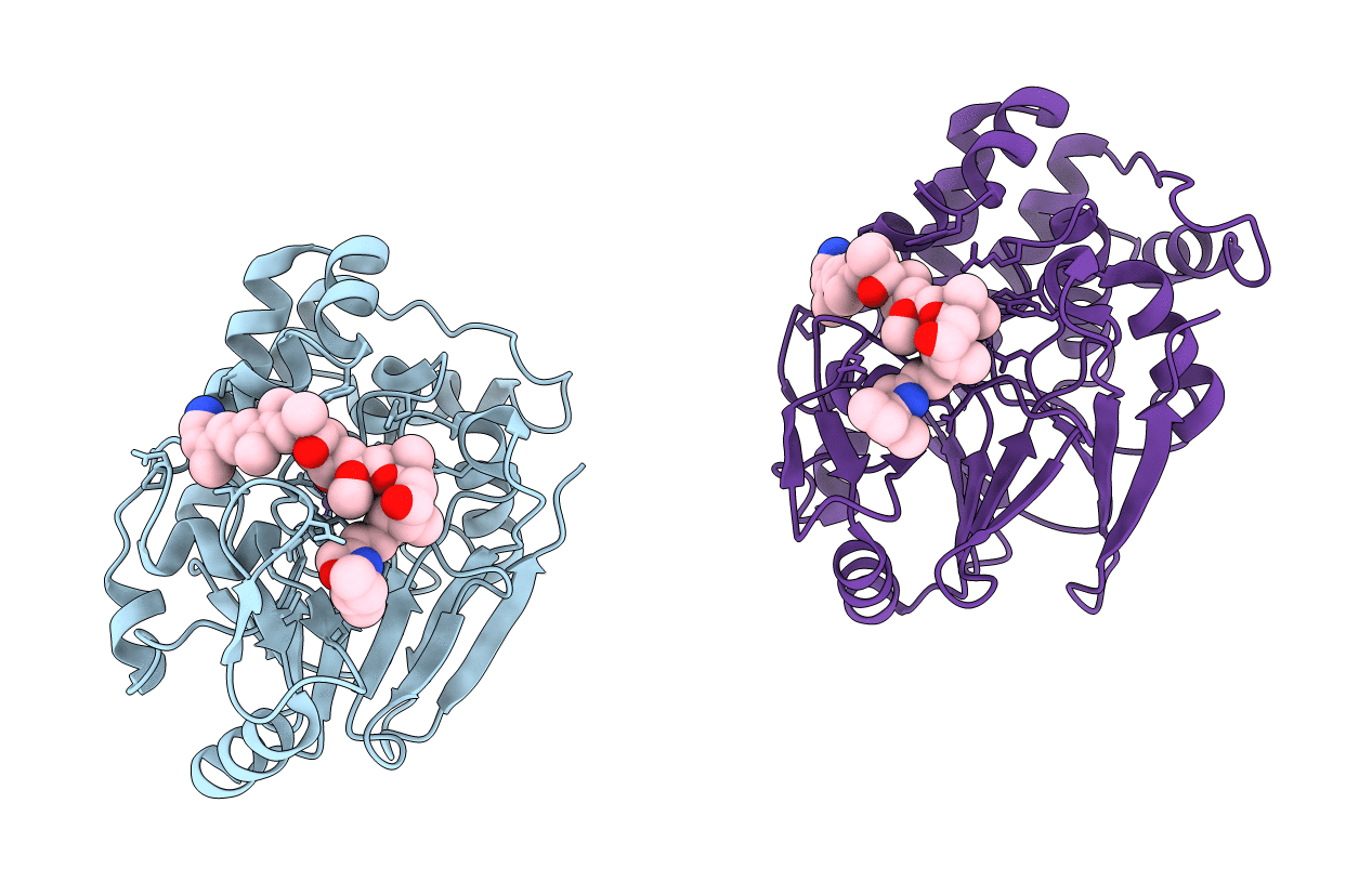
Title:
CRYSTAL STRUCTURE OF THE COMPLEX BETWEEN CALYCULIN A AND THE CATALYTIC SUBUNIT OF PROTEIN PHOSPHATASE 1
Biological Source:
Source Organism(s):
Homo sapiens (Taxon ID: 9606)
Expression System(s):
Method Details:
Experimental Method:
Resolution:
2.00 Å
R-Value Free:
0.21
R-Value Work:
0.18
Space Group:
P 21 21 2
