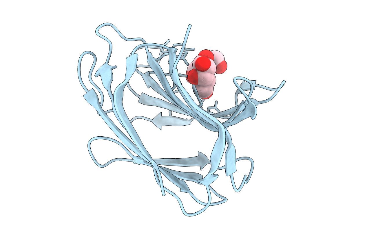
1IS4 image
Deposition Date 2001-11-12
Release Date 2002-09-18
Last Version Date 2024-04-03
Entry Detail
PDB ID:
1IS4
Title:
LACTOSE-LIGANDED CONGERIN II
Biological Source:
Source Organism:
Host Organism:
Method Details:
Experimental Method:
Resolution:
1.90 Å
R-Value Free:
0.21
R-Value Work:
0.16
R-Value Observed:
0.16
Space Group:
P 42 21 2
Macromolecular Entities
Polymer Type:polypeptide(L)
Molecule:CONGERIN II
Chain IDs:A
Chain Length:135
Number of Molecules:1
Biological Source:Conger myriaster
Peptide-like Molecules
PRD_900004
Primary Citation
Crystal Structure of a Conger Eel Galectin (Congerin II) at 1.45 A Resolution: Implication for the Accelerated Evolution of a New Ligand-Binding Site Following Gene Duplication
J.Mol.Biol. 321 879 889 (2002)
PMID: 12206768 DOI: 10.1016/S0022-2836(02)00700-3

Abstact

The crystal structure of congerin II, a galectin family lectin from conger eel, was determined at 1.45A resolution. The previously determined structure of its isoform, congerin I, had revealed a fold evolution via strand swap; however, the structure of congerin II described here resembles other prototype galectins. A comparison of the two congerin genes with that of several other galectins suggests acceralated evolution of both congerin genes following gene duplication. The presence of a Mes (2-[N-morpholino]ethanesulfonic acid) molecule near the carbohydrate-binding site in the crystal structure points to the possibility of an additional binding site in congerin II. The binding site consists of a group of residues that had been replaced following gene duplication suggesting that the binding site was built under selective pressure. Congerin II may be a protein specialized for biological defense with an affinity for target carbohydrates on parasites' cell surface.

Legend

Protein

Chemical

Disease

Primary Citation of related structures