1IR1 image
Deposition Date 2001-08-31
Release Date 2002-03-13
Last Version Date 2023-11-15
Entry Detail
PDB ID:
1IR1
Keywords:
Title:
Crystal Structure of Spinach Ribulose-1,5-Bisphosphate Carboxylase/Oxygenase (Rubisco) Complexed with CO2, Mg2+ and 2-Carboxyarabinitol-1,5-Bisphosphate
Biological Source:
Source Organism:
Method Details:
Experimental Method:
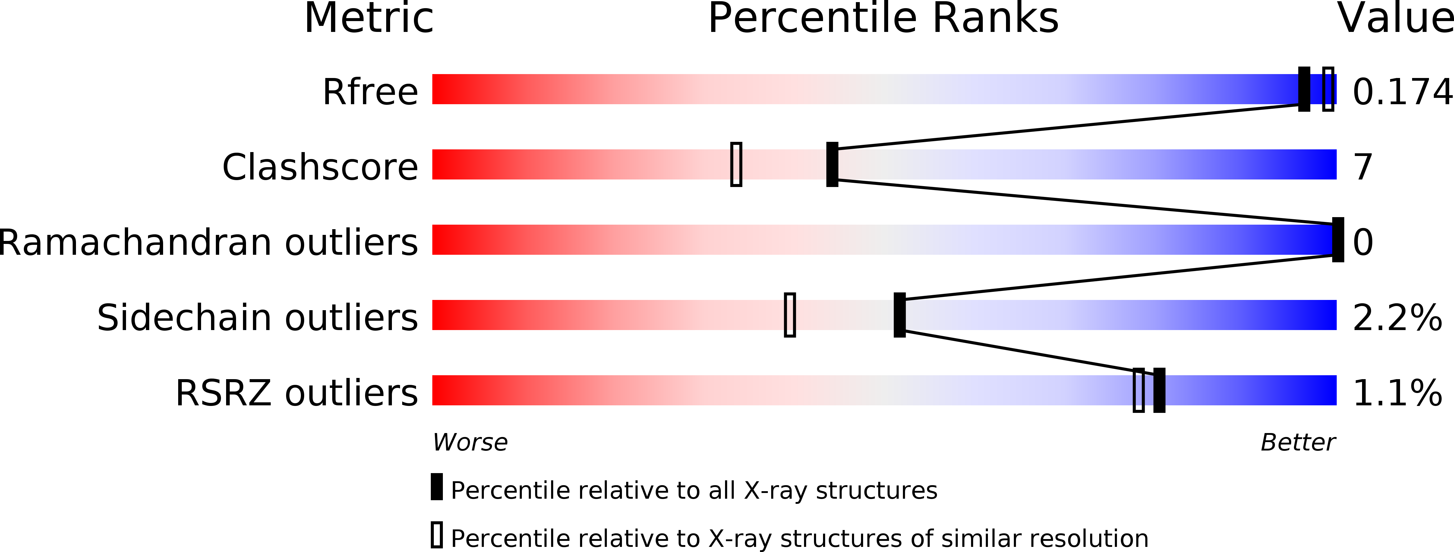
Resolution:
1.80 Å
R-Value Free:
0.17
R-Value Work:
0.15
Space Group:
C 2 2 21
Macromolecular Entities
Polymer Type:polypeptide(L)
Molecule:Large subunit of Rubisco
Gene (Uniprot):rbcL
Chain IDs:A, C (auth: B), E (auth: C), G (auth: D)
Chain Length:475
Number of Molecules:4
Biological Source:Spinacia oleracea
Polymer Type:polypeptide(L)
Molecule:Small subunit of Rubisco
Gene (Uniprot):RBCS2
Chain IDs:B (auth: S), D (auth: T), F (auth: U), H (auth: V)
Chain Length:123
Number of Molecules:4
Biological Source:Spinacia oleracea
Modified Residue
Compound ID Chain ID Parent Comp ID Details 2D Image
KCX A LYS LYSINE NZ-CARBOXYLIC ACID
MME B MET N-METHYL METHIONINE
Primary Citation
Crystal structure of activated ribulose-1,5-bisphosphate carboxylase/oxygenase from green alga Chlamydomonas reinhardtii complexed with 2-carboxyarabinitol-1,5-bisphosphate.
J.Mol.Biol. 316 679 691 (2002)
PMID: 11866526 DOI: 10.1006/jmbi.2001.5381

Abstact

Ribulose-1,5-bisphosphate carboxylase/oxygenase (rubisco) catalyzes the initial steps of photosynthetic carbon reduction and photorespiratory carbon oxidation cycles by combining CO(2) and O(2), respectively, with ribulose-1,5-bisphosphate. Many photosynthetic organisms have form I rubiscos comprised of eight large (L) and eight small (S) subunits. The crystal structure of the complex of activated rubisco from the green alga Chlamydomonas reinhardtii and the reaction intermediate analogue 2-carboxyarabinitol-1,5-bisphosphate (2-CABP) has been solved at 1.84 A resolution (R(cryst) of 15.2 % and R(free) of 18.1 %). The subunit arrangement of Chlamydomonas rubisco is the same as those of the previously solved form I rubiscos. Especially, the present structure is very similar to the activated spinach structure complexed with 2-CABP in the L-subunit folding and active-site conformation, but differs in S-subunit folding. The central insertion of the Chlamydomonas S-subunit forms the longer betaA-betaB loop that protrudes deeper into the solvent channel of rubisco than higher plant, cyanobacterial, and red algal (red-like) betaA-betaB loops. The C-terminal extension of the Chlamydomonas S-subunit does not protrude into the solvent channel, unlike that of the red algal S-subunit, but lies on the protein surface anchored by interactions with the N-terminal region of the S-subunit. Further, the present high-resolution structure has revealed novel post-translational modifications. Residue 1 of the S-subunit is N(alpha)-methylmethionine, residues 104 and 151 of the L-subunit are 4-hydroxyproline, and residues 256 and 369 of the L-subunit are S(gamma)-methylcysteine. Furthermore, the unusual electron density of residue 471 of the L-subunit, which has been deduced to be threonine from the genomic DNA sequence, suggests that the residue is isoleucine produced by RNA editing or O(gamma)-methylthreonine.

Legend

Protein

Chemical

Disease

Primary Citation of related structures