1IOU image
Deposition Date 2001-04-09
Release Date 2003-05-06
Last Version Date 2023-12-27
Entry Detail
PDB ID:
1IOU
Title:
SOLUTION STRUCTURE OF YKT6P (1-140)
Biological Source:
Source Organism:
Host Organism:
Method Details:
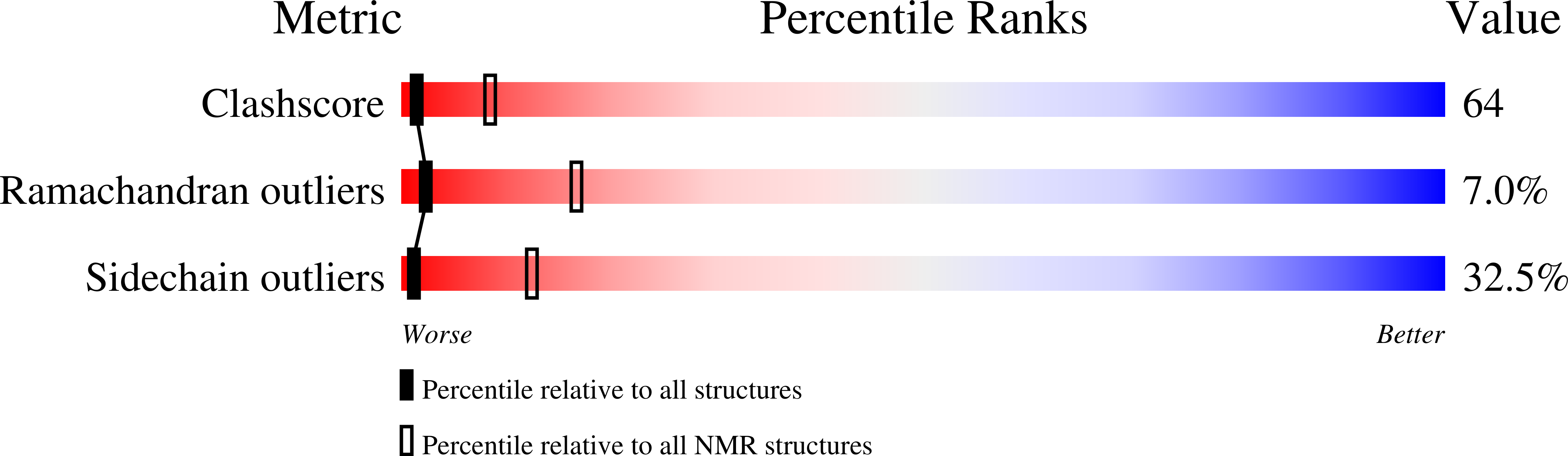
Experimental Method:
Conformers Calculated:
200
Conformers Submitted:
20
Selection Criteria:
structures with the least restraint violations,structures with the lowest energy
Macromolecular Entities
Structures with similar UniProt ID
Protein Blast
Polymer Type:polypeptide(L)
Molecule:YKT6P
Gene (Uniprot):YKT6
Chain IDs:A
Chain Length:140
Number of Molecules:1
Biological Source:Saccharomyces cerevisiae
Ligand Molecules
Primary Citation
An autoinhibitory mechanism for nonsyntaxin SNARE proteins revealed by the structure of Ykt6p
SCIENCE 293 698 702 (2001)
PMID: 11474112 DOI: 10.1126/science.1062950

Abstact

Ykt6p is a nonsyntaxin SNARE implicated in multiple intracellular membrane trafficking steps. Here we present the structure of the NH2-terminal domain of Ykt6p (Ykt6pN, residues 1 to 140). The structure of Ykt6pN differed entirely from that of syntaxin and resembled the overall fold of the actin regulatory protein, profilin. Like some syntaxins, Ykt6p adopted a folded back conformation in which Ykt6pN bound to its COOH-terminal core domain. The NH2-terminal domain plays an important biological role in the function of Ykt6p, which in vitro studies revealed to include influencing the kinetics and proper assembly of SNARE complexes.

Legend

Protein

Chemical

Disease

Primary Citation of related structures
Feedback Form
Name
Email
Institute
Feedback