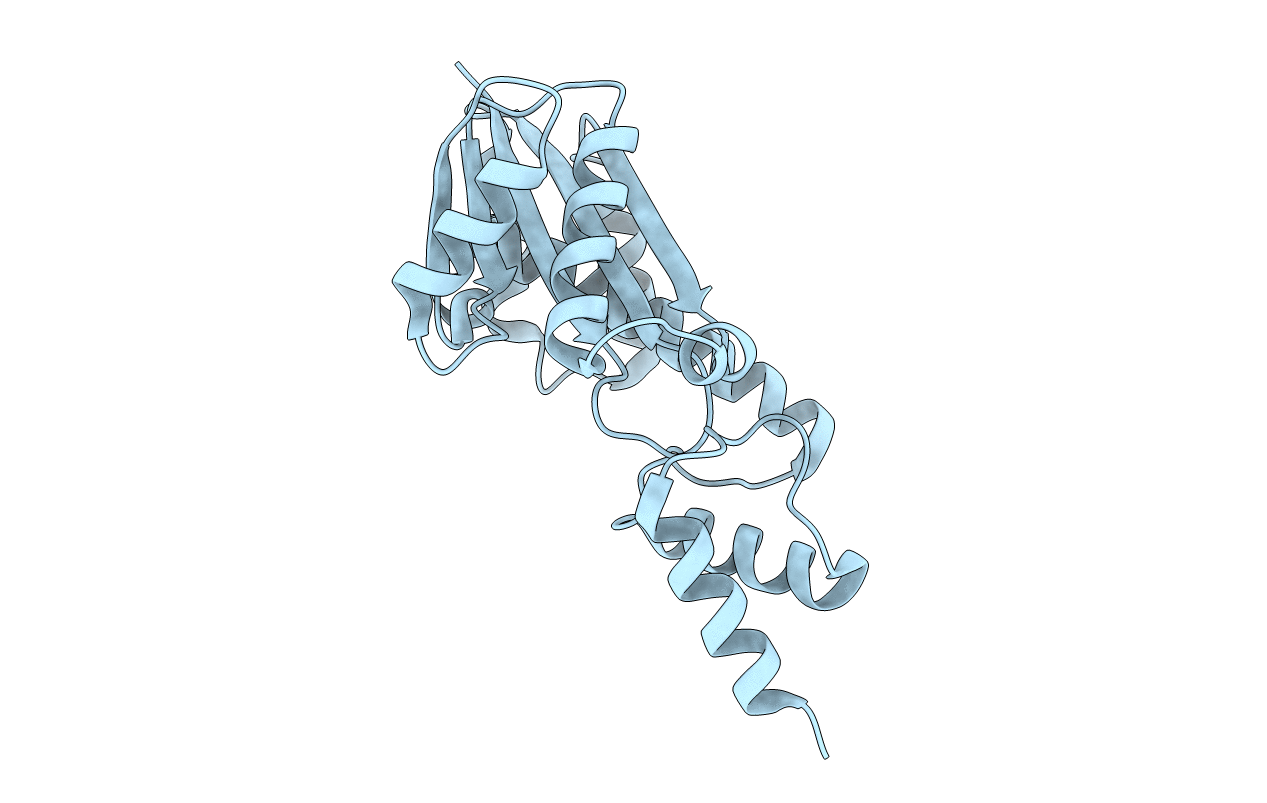
1IO2 image
Deposition Date 2000-12-28
Release Date 2001-04-18
Last Version Date 2023-12-27
Entry Detail
PDB ID:
1IO2
Keywords:
Title:
Crystal structure of type 2 ribonuclease h from hyperthermophilic archaeon, thermococcus kodakaraensis kod1
Biological Source:
Source Organism:
Host Organism:
Method Details:
Experimental Method:
Resolution:
2.00 Å
R-Value Free:
0.27
R-Value Work:
0.22
R-Value Observed:
0.22
Space Group:
P 21 21 21
Macromolecular Entities
Structures with similar UniProt ID
Protein Blast
Polymer Type:polypeptide(L)
Molecule:RIBONUCLEASE HII
Gene (Uniprot):rnhB
Chain IDs:A
Chain Length:213
Number of Molecules:1
Biological Source:Thermococcus kodakarensis
Primary Citation
Catalytic center of an archaeal type 2 ribonuclease H as revealed by X-ray crystallographic and mutational analyses.
Protein Sci. 10 707 714 (2001)
PMID: 11274461 DOI: 10.1110/ps.48001

Abstact

The catalytic center of an archaeal Type 2 RNase H has been identified by a combination of X-ray crystallographic and mutational analyses. The crystal structure of the Type 2 RNase H from Thermococcus kodakaraensis KOD1 has revealed that the N-terminal major domain adopts the RNase H fold, despite the poor sequence similarity to the Type 1 RNase H. Mutational analyses showed that the catalytic reaction requires four acidic residues, which are well conserved in the Type 1 RNase H and the members of the polynucleotidyl transferase family. Thus, the Type 1 and Type 2 RNases H seem to share a common catalytic mechanism, except for the requirement of histidine as a general base in the former enzyme. Combined with the results from deletion mutant analyses, the structure suggests that the C-terminal domain of the Type 2 RNase H is involved in the interaction with the DNA/RNA hybrid.

Legend

Protein

Chemical

Disease

Primary Citation of related structures