
Deposition Date
2001-05-14
Release Date
2002-03-20
Last Version Date
2024-11-13
Entry Detail
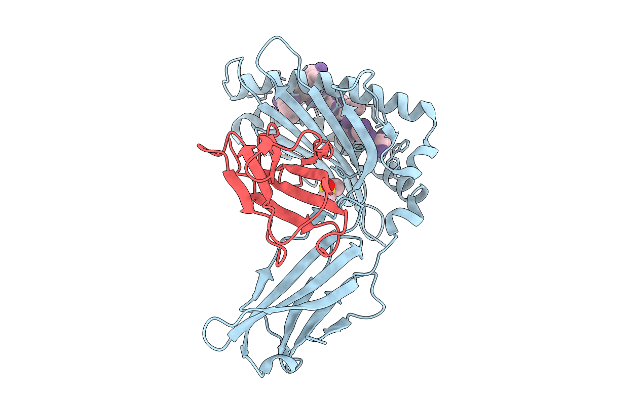
PDB ID:
1INQ
Keywords:
Title:
Structure of Minor Histocompatibility Antigen peptide, H13a, complexed to H2-Db
Biological Source:
Source Organism(s):
Mus musculus (Taxon ID: 10090)
Expression System(s):
Method Details:
Experimental Method:
Resolution:
2.20 Å
R-Value Free:
0.25
R-Value Work:
0.20
R-Value Observed:
0.21
Space Group:
C 1 2 1
