Deposition Date
2001-05-11
Release Date
2001-10-03
Last Version Date
2024-05-22
Entry Detail
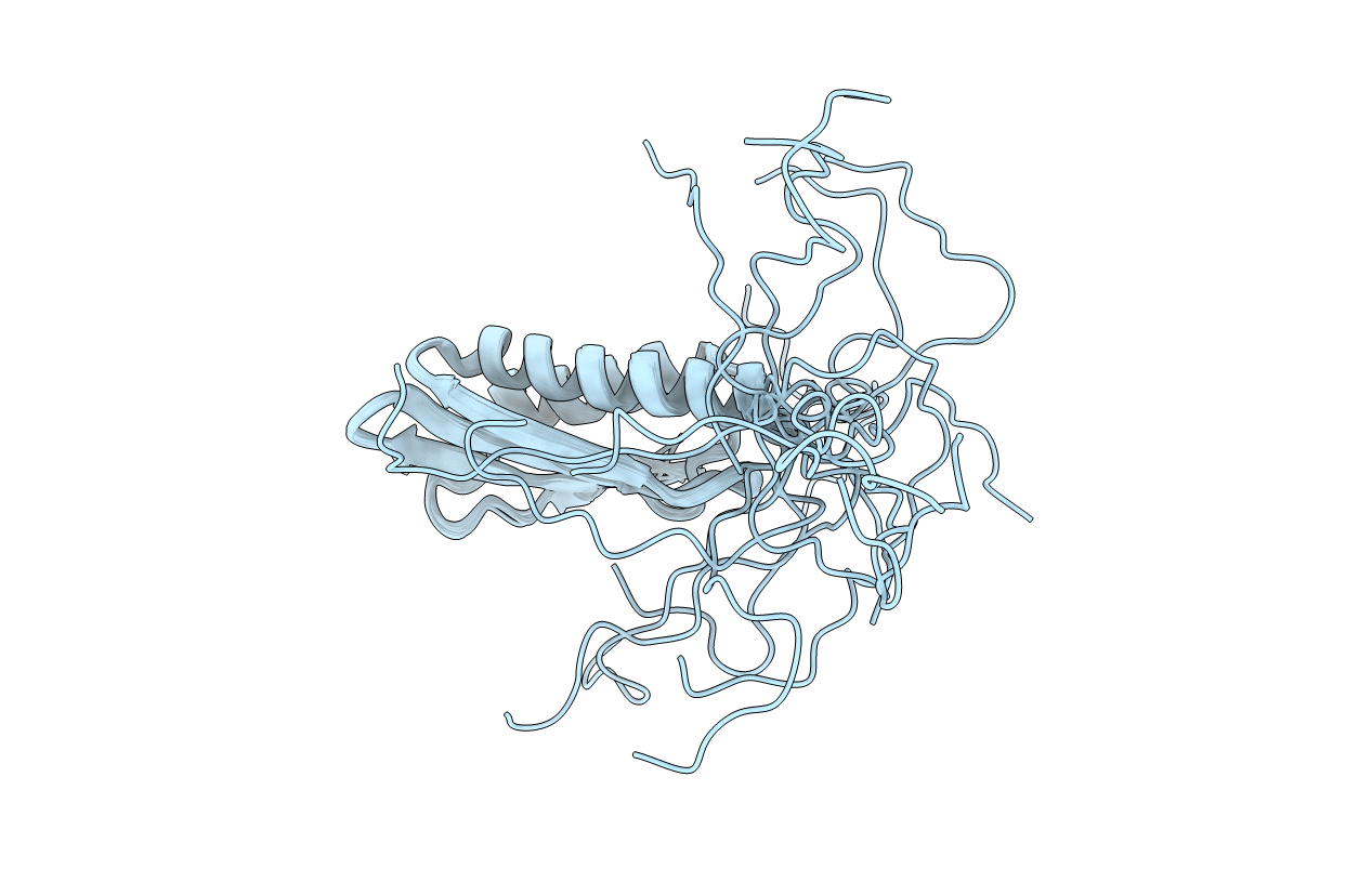
PDB ID:
1IMU
Keywords:
Title:
Solution Structure of HI0257, a Ribosome Binding Protein
Biological Source:
Source Organism(s):
Haemophilus influenzae (Taxon ID: 727)
Expression System(s):
Method Details:
Experimental Method:
Conformers Calculated:
39
Conformers Submitted:
20
Selection Criteria:
structures with the least restraint violations, structures with the lowest energy