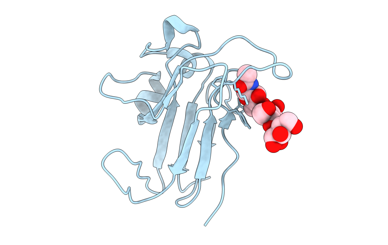
1IKO image
Deposition Date 2001-05-03
Release Date 2002-05-03
Last Version Date 2024-11-06
Entry Detail
PDB ID:
1IKO
Title:
CRYSTAL STRUCTURE OF THE MURINE EPHRIN-B2 ECTODOMAIN
Biological Source:
Source Organism:
Mus musculus (Taxon ID: 10090)
Host Organism:
Method Details:
Experimental Method:
Resolution:
1.92 Å
R-Value Free:
0.24
R-Value Work:
0.21
R-Value Observed:
0.21
Space Group:
P 65 2 2
Macromolecular Entities
Structures with similar UniProt ID
Protein Blast
Polymer Type:polypeptide(L)
Molecule:EPHRIN-B2
Gene (Uniprot):Efnb2
Chain IDs:A (auth: P)
Chain Length:178
Number of Molecules:1
Biological Source:Mus musculus
Modified Residue
Compound ID Chain ID Parent Comp ID Details 2D Image
ASN A ASN GLYCOSYLATION SITE
Primary Citation
Crystal structure of an ephrin ectodomain.
Dev.Cell 1 83 92 (2001)
PMID: 11703926 DOI: 10.1016/S1534-5807(01)00002-8

Abstact

Eph receptor tyrosine kinases and their membrane-associated ligands, the ephrins, are essential regulators of axon guidance, cell migration, segmentation, and angiogenesis. There are two classes of vertebrate ephrin ligands which have distinct binding specificities for their cognate receptors. Multimerization of the ligands is required for receptor activation, and ephrin ligands themselves signal intracellularly upon binding Eph receptors. We have determined the structure of the extracellular domain of mouse ephrin-B2. The ephrin ectodomain is an eight-stranded beta barrel with topological similarity to plant nodulins and phytocyanins. Based on the structure, we have identified potential surface determinants of Eph/ephrin binding specificity and a ligand dimerization region. The high sequence similarity among ephrin ectodomains indicates that all ephrins may be modeled upon the ephrin-B2 structure presented here.

Legend

Protein

Chemical

Disease

Primary Citation of related structures