1IK6 image
Entry Detail
PDB ID:
1IK6
Keywords:
Title:
3D structure of the E1beta subunit of pyruvate dehydrogenase from the archeon Pyrobaculum aerophilum
Biological Source:
Source Organism:
PDB Version:
Deposition Date:
2001-05-02
Release Date:
2001-12-19
Method Details:
Experimental Method:
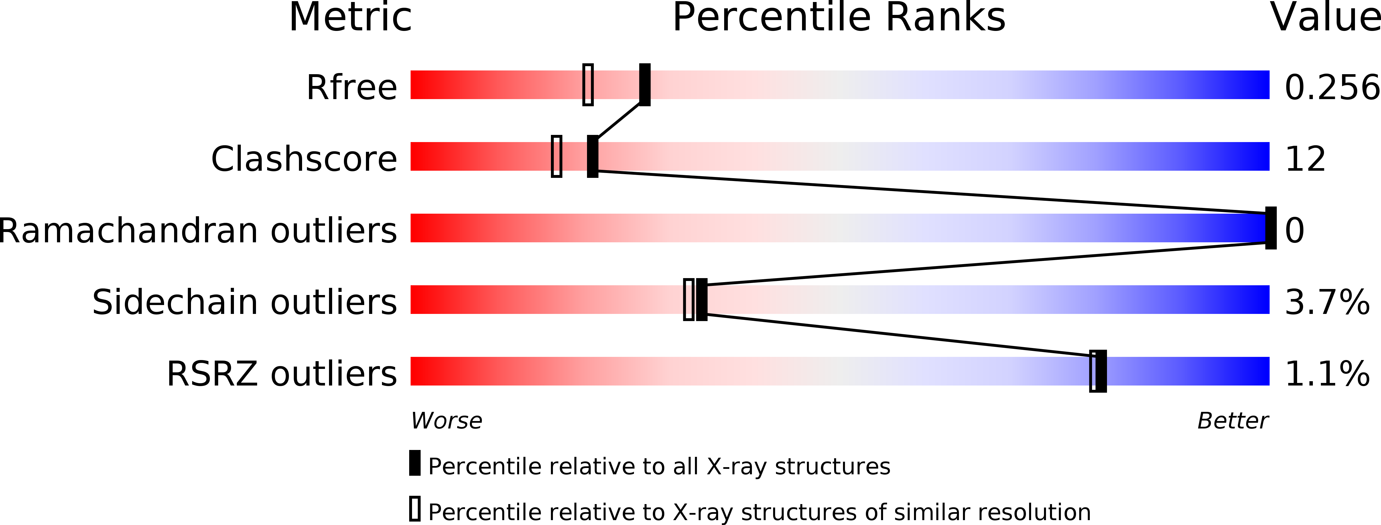
Resolution:
2.00 Å
R-Value Free:
0.25
R-Value Work:
0.21
Space Group:
I 2 2 2
Macromolecular Entities
Polymer Type:polypeptide(L)
Description:pyruvate dehydrogenase
Chain IDs:A
Chain Length:369
Number of Molecules:1
Biological Source:Pyrobaculum aerophilum
Primary Citation
3D structure and significance of the GPhiXXG helix packing motif in tetramers of the E1beta subunit of pyruvate dehydrogenase from the archeon Pyrobaculum aerophilum.
Biochemistry 40 14484 14492 (2001)
PMID: 11724561 DOI: 10.1021/bi011016k

Abstact

As part of a structural genomics project, we have determined the 2.0 A structure of the E1beta subunit of pyruvate dehydrogenase from Pyrobaculum aerophilum (PA), a thermophilic archaeon. The overall fold of E1beta from PA is closely similar to the previously determined E1beta structures from humans (HU) and P. putida (PP). However, unlike the HU and PP structures, the PA structure was determined in the absence of its partner subunit, E1alpha. Significant structural rearrangements occur in E1beta when its E1alpha partner is absent, including rearrangement of several secondary structure elements such as helix C. Helix C is buried by E1alpha in the HU and PP structures, but makes crystal contacts in the PA structure that lead to an apparent beta(4) tetramer. Static light scattering and sedimentation velocity data are consistent with the formation of PA E1beta tetramers in solution. The interaction of helix C with its symmetry-related counterpart stabilizes the tetrameric interface, where two glycine residues on the same face of one helix create a packing surface for the other helix. This GPhiXXG helix-helix interaction motif has previously been found in interacting transmembrane helices, and is found here at the E1alpha-E1beta interface for both the HU and PP alpha(2)beta(2) tetramers. As a case study in structural genomics, this work illustrates that comparative analysis of protein structures can identify the structural significance of a sequence motif.

Legend

Protein

Chemical

Disease

Primary Citation of related structures