1IJL image
Deposition Date 2001-04-27
Release Date 2001-12-28
Last Version Date 2024-10-16
Entry Detail
PDB ID:
1IJL
Keywords:
Title:
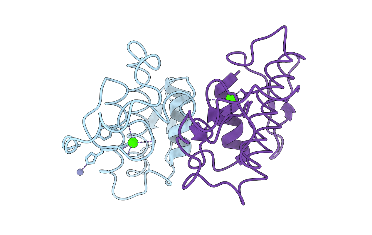
Crystal structure of acidic phospholipase A2 from deinagkistrodon acutus
Biological Source:
Source Organism:
Method Details:
Experimental Method:
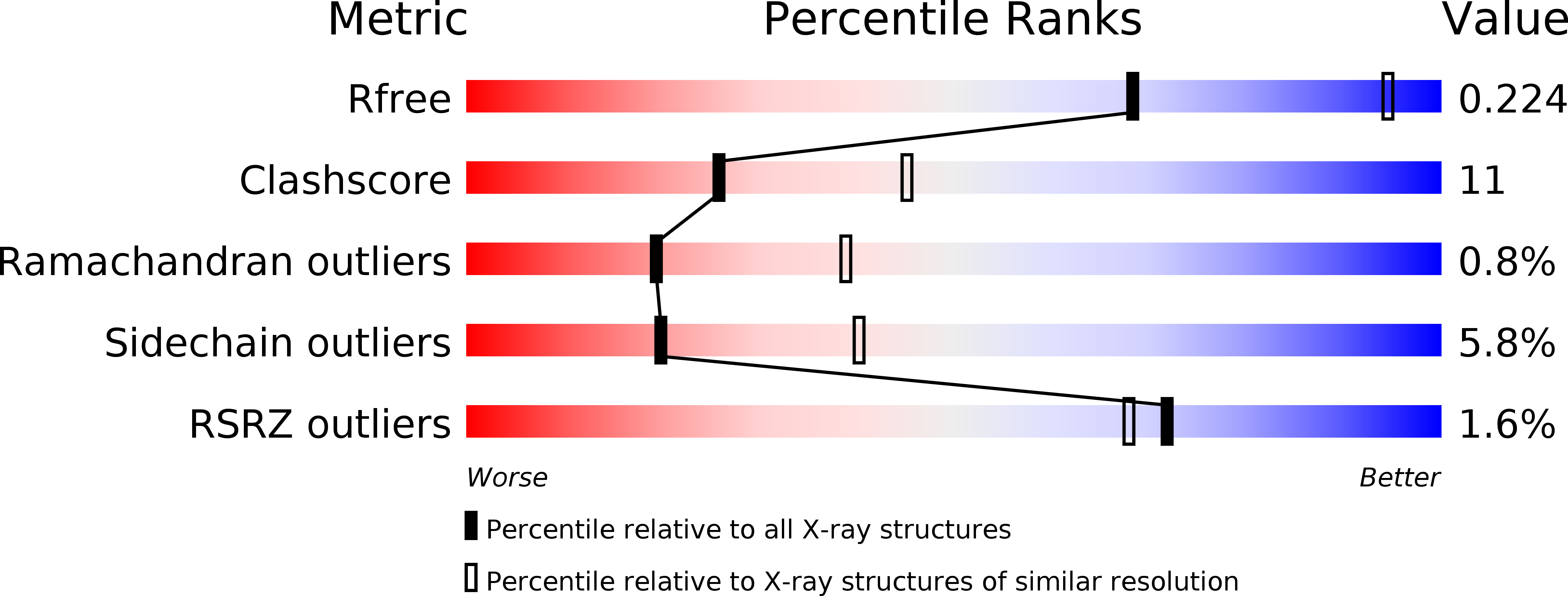
Resolution:
2.60 Å
R-Value Free:
0.22
R-Value Work:
0.18
Space Group:
P 1 21 1
Macromolecular Entities
Polymer Type:polypeptide(L)
Molecule:PHOSPHOLIPASE A2
Chain IDs:A, B
Chain Length:123
Number of Molecules:2
Biological Source:Deinagkistrodon acutus
Primary Citation
Structure of an acidic phospholipase A2 from the venom of Deinagkistrodon acutus.
Acta Crystallogr.,Sect.D 58 104 110 (2002)
PMID: 11752784 DOI: 10.1107/S0907444901018170

Abstact

An acidic phospholipase A(2) was purified from Deinagkistrodon acutus (Agkistrodon acutus) which displays an inhibitory effect on platelet aggregation. The three-dimensional structure of the enzyme was determined by molecular replacement at 2.6 A resolution with a crystallographic R factor of 18.40% (R(free) = 22.50%) and reasonable stereochemistry. Two molecules in the asymmetric unit form a dimer and the dimer formation accompanies a significant conformational adaptation of segment 14-23, a constituent of the 'interface recognition site' (IRS). This probably reflects the inherent structural flexibility of the IRS. The possible expansion of the site for inhibiting platelet aggregation as proposed previously [Wang et al. (1996), J. Mol. Biol. 255, 669-676] is discussed.

Legend

Protein

Chemical

Disease

Primary Citation of related structures