1IJJ image
Entry Detail
PDB ID:
1IJJ
Title:
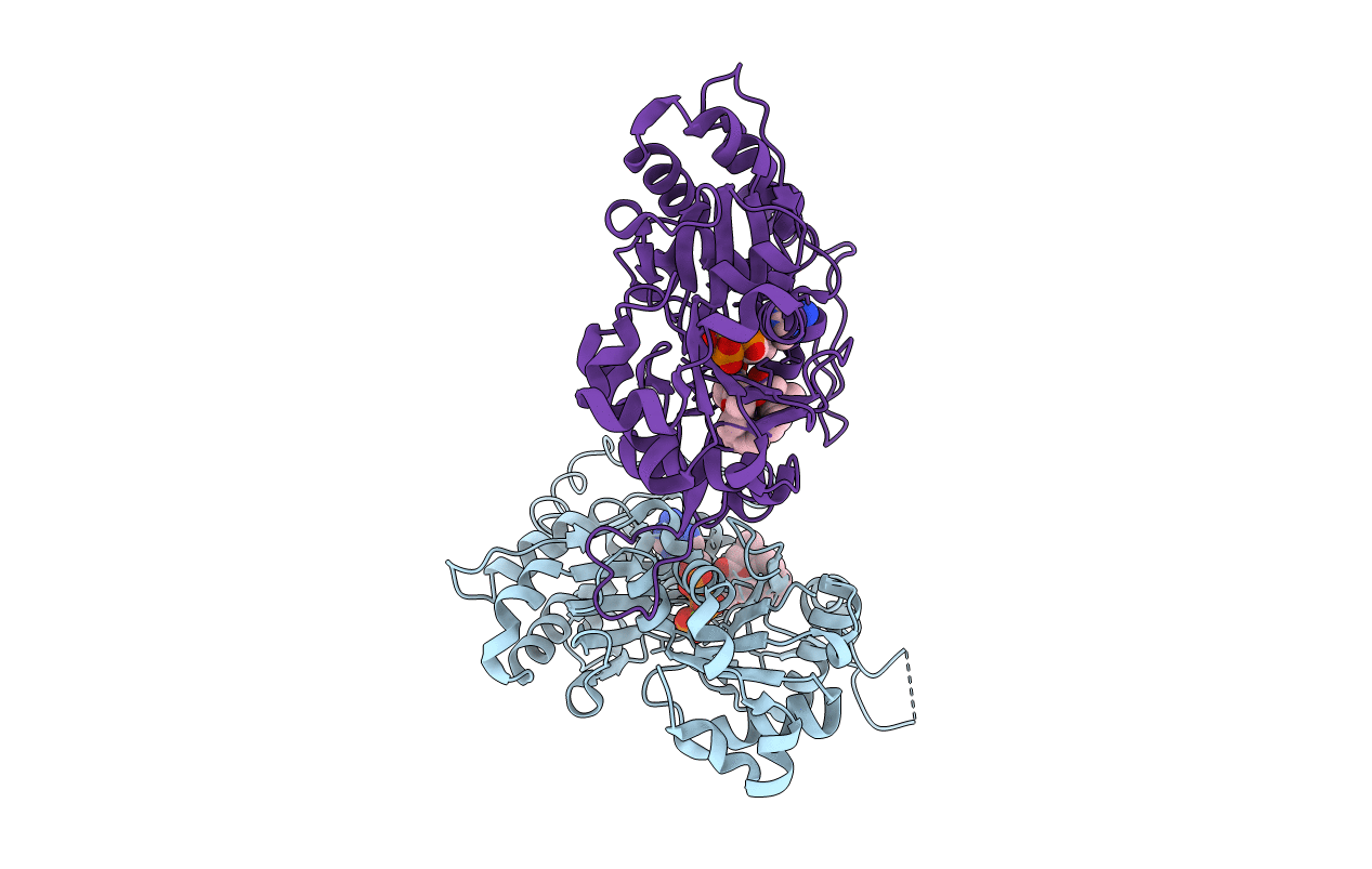
THE X-RAY CRYSTAL STRUCTURE OF THE COMPLEX BETWEEN RABBIT SKELETAL MUSCLE ACTIN AND LATRUNCULIN A AT 2.85 A RESOLUTION
Biological Source:
Source Organism:
PDB Version:
Deposition Date:
2001-04-26
Release Date:
2002-04-15
Method Details:
Experimental Method:
Resolution:
2.85 Å
R-Value Free:
0.30
R-Value Work:
0.23
Space Group:
P 21 21 21
Macromolecular Entities
Polymer Type:polypeptide(L)
Description:ACTIN, ALPHA SKELETAL MUSCLE
Chain IDs:A, B
Chain Length:377
Number of Molecules:2
Biological Source:Oryctolagus cuniculus
Primary Citation
Polylysine induces an antiparallel actin dimer that nucleates filament assembly: crystal structure at 3.5-A resolution
J.Biol.Chem. 277 20999 21006 (2002)
PMID: 11932258 DOI: 10.1074/jbc.M201371200

Abstact

An antiparallel actin dimer has been proposed to be an intermediate species during actin filament nucleation. We now show that latrunculin A, a marine natural product that inhibits actin polymerization, arrests polylysine-induced nucleation at the level of an antiparallel dimer, resulting in its accumulation. These dimers, when composed of pyrene-labeled actin subunits, give rise to a fluorescent excimer, permitting detection during polymerization in vitro. We report the crystallographic structure of the polylysine-actin-latrunculin A complex at 3.5-A resolution. The non-crystallographic contact is consistent with a dimeric structure and confirms the antiparallel orientation of its subunits. The crystallographic contacts reveal that the mobile DNase I binding loop of one subunit of a symmetry-related antiparallel actin dimer is partially stabilized in the interface between the two subunits of a second antiparallel dimer. These results provide a potential explanation for the paradoxical nucleation of actin filaments that have exclusively parallel subunits by a dimer containing antiparallel subunits.

Legend

Protein

Chemical

Disease

Primary Citation of related structures