1IIJ image
Deposition Date 2001-04-23
Release Date 2001-06-27
Last Version Date 2024-05-22
Entry Detail
PDB ID:
1IIJ
Title:
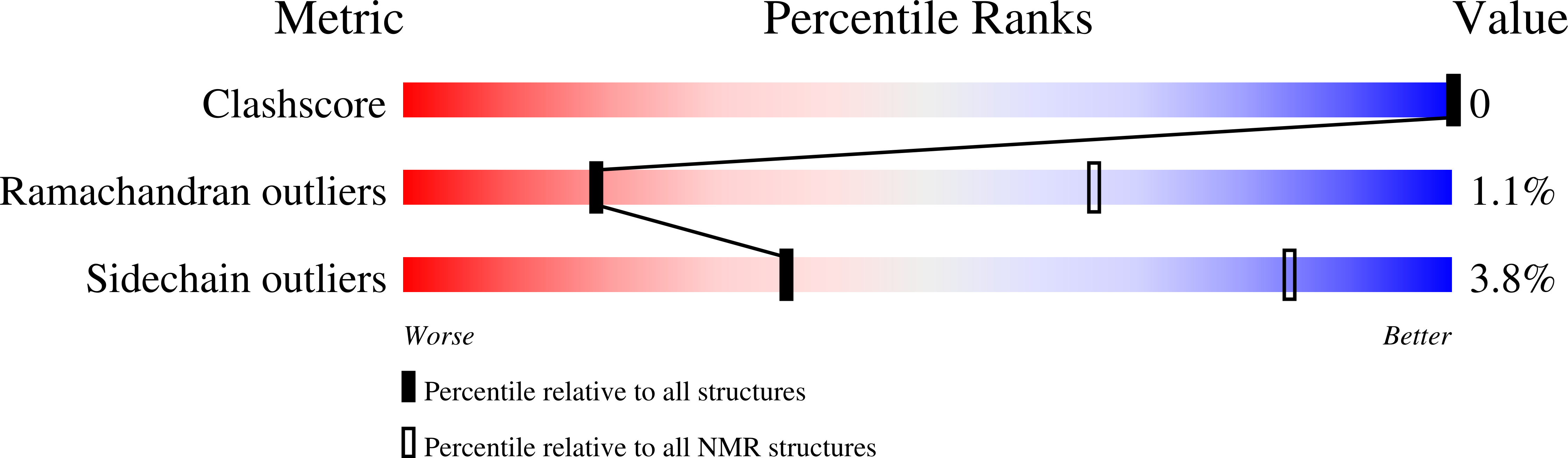
SOLUTION STRUCTURE OF THE NEU/ERBB-2 MEMBRANE SPANNING SEGMENT
Biological Source:
Source Organism:
Method Details:
Experimental Method:
Conformers Calculated:
20
Conformers Submitted:
5
Selection Criteria:
structures with the lowest energy
Macromolecular Entities
Structures with similar UniProt ID
Protein Blast
Polymer Type:polypeptide(L)
Molecule:ERBB-2 RECEPTOR PROTEIN-TYROSINE KINASE
Gene (Uniprot):Erbb2
Chain IDs:A
Chain Length:35
Number of Molecules:1
Biological Source:
Ligand Molecules
Primary Citation
Evidence for an alpha-helix --> pi-bulge helicity modulation for the neu/erbB-2 membrane-spanning segment. A 1H NMR and circular dichroism study.
Biochemistry 40 6534 6540 (2001)
PMID: 11371217 DOI: 10.1021/bi0027938

Abstact

The 35-residue peptide corresponding to the very hydrophobic transmembrane region of the tyrosine kinase receptor neu, Neu(TM35), has been synthesized. The peptide can be solubilized in millimolar concentrations in TFE or incorporated into an SDS-water micellar solution or into well-hydrated DMPC/DCPC bicelles. In all these media, circular dichroism demonstrated that the peptide adopts a helical structure for about 80% of its amino acids. The peptide is monomeric below 2 mM in TFE, as also determined by variable concentration experiments. The three-dimensional solution structure in TFE has been obtained by homonuclear proton NMR and shows a well-defined alpha-helix from residues 4 to 21, then a pi-bulge from Ile(22) to Gly(28), and a final short alpha-helix from positions 29 to 32. This experimental finding is in agreement with structures predicted recently by molecular dynamics calculations in a vacuum [Sajot, N., and Genest, M. (2000) Eur. Biophys. J. 28, 648-662]. The biological implications of a possible retention of this structure in a membrane environment are finally discussed.

Legend

Protein

Chemical

Disease

Primary Citation of related structures